Organ Donations After Cardiac Death Soar in US, Expand Transplant Lifeline 

One of the significant changes in the way people approach organ donation in the United States is the growing availability of transplantation organs, with almost half of all donors being patients whose heart has gone dead, according to latest studies.

According to the study by scientists at NYU Langone Health, it has been established that donation after circulatory death (DCD) has increased significantly in the last 25 years – marking an increase of 2 percent of all donors in 2000 to 49 percent in 2025. According to the findings published in Journal of the American Medicine, the development of medical technology is transforming transplant medicine.

The growth has been realized when demand is acute. According to the United Network for Organ Sharing, more than 100,000 individuals are already on transplant waiting lists in the U.S., and this fact requires finding new sources of viable organs.

Conventionally, organs donated have been infected out of patients who have been declared brain dead, those organs keep being oxygenated with the heart still beating. Conversely, DCD deals with patients who are not yet dead, but are on life support. In case life-sustaining treatment is withdrawn and the patient dies in a given period, then organs can be removed to be transplanted, though it must be otherwise previously agreed.

Drawbacks Overcome With Tech 

In past, organs transplanted by such sources were less viable because of a short period of lack of oxygen following the cessation of the heart. Nevertheless, these drawbacks have been overcome with the recent technology advances.

Improved organ preservation has been achieved using techniques like normothermic regional perfusion in which blood flow to organs is resumed following cardiac death and machine perfusion systems in which oxygenated fluids are delivered extravascularly. These inventions have made innovations through which the surgeons can safely utilize organs that were not considered to be perfect.

According to researchers, this has expanded the pool of donors. The researchers discovered that current DCD donors are older individuals with higher probabilities of underlying diseases like diabetes or hypertension as compared to previous generation, which is more inclusive in the selection of the donor.

Syed Ali Husain, the lead author, indicated that the increase in circulatory-death donations is already producing a tangible impact, and thousands of patients were already getting transplants who otherwise would not have been able to survive the wait.

Regional Disparity Persists

The national data on transplants also indicated that there were disparities in the connections of the regions. DCD donors contributed up to 73 per cent of all donations in certain regions of the country and only 11 per cent in other regions indicating a lack of balance in the practice.

The researchers working on the study underlined the importance of developing uniform national standards and ongoing involvement of the population to protect the ethics and preserve a trusting attitude towards the process of donating.

Researchers believe that more papers are required to understand long-term outcomes and enhance protocols as the DCD is becoming more popular. Further research will aim to enhance the process of donor identification and understand the performance of organs of donors who died of a circulatory death as opposed to the performance of organs of those who died of a traditional brain-death.

The results represent an important development in the field of transplant medicine – one that may aid in reducing the disparity between supply and demand of organs, and also pose new challenges to clinical practice, ethics and popular opinion.

Also Read:

New Chip Helps Diagnose Heart Attacks Based on Blood Test in Minutes

Heart attack prevention lags for people with stroke, peripheral artery disease: Study

New strategy prevents pancreatic cancer removing microscopic lesions early

A recent preclinical trial using mice reveals that the precancerous cells of the pancreas can be disposed before they can develop into a tumor. Application of an experimental therapy to attack microscopic precancerous lesions in the pancreas was shown to increase survival in mouse models of a pancreatic ductal adenocarcinoma (PDAC) by nearly 2 times even when the same therapy was administered when the cancer was already present.

The study was carried out under the leadership of physician-scientists of the Perelman school of medicine in the University of Pennsylvania and Abramson cancer center of Penn medicine, which is released to date in Science.

It is also the first instance that scientists have demonstrated that a medical intervention could prevent growth of pre-cancerous lesions in the pancreas before it becomes pancreatic cancer and this is a good indication to the growing area of cancer interception.

I believe that the next age of cancer treatment will be interception, and I am confident that such an endeavor will be developed by cancer treatment experts like Robert Vonderheide, the director of the Abramson Cancer Center. Pancreatic cancer has had a stubborn poor prognosis, scarce treatment options as well as no tested screening and prevention measures. And should we be able to get a means of intercepting it–of detecting and counteracting abnormalities in their first beginnings of malignancy, it would be an issue game changer.

Cancer interception is different to cancer prevention.

Contrary to prevention approaches, like HPV vaccination or ceasing a smoking habit, which seek to ensure that the cancer never develops at all, cancer interception focuses on the first stages of a cell becoming malignant. Colonoscopy can be taken to illustrate an instance of a mechanical interception where the precancerous polyps are removed before they turn into colorectal cancer. Since the harder the malignancy is to treat, the larger it is, the idea of curing the malignant growths before they become cancer is quite a logical one in theory, yet challenging to be proven.

“The paper is a preclinical evidence-of-concept demonstrating that medical cancer interception is better than treatment of a diagnosis,” said the lead author, Minh Than, a clinical and research fellow in the field of Hematology-Oncology. “This research demonstrates the strength of using a proactive approach to cancer, as opposed to a reactive one. This will be interesting to test in our patients in the second stage of this work.”

RAS inhibition is an efficient way of intercepting cancer in mice.

In this research, the researchers employed two experimental inhibitors that are directed to the cancer causing gene, KRAS. KRAS mutations that cause more than 90 percent of pancreatic cancers are the most frequent cancer-causing gene mutation found in all cancers and has been traditionally thought to be undruggable.

The first KRAS inhibitor was approved in 2021 to treat non-small cell lung cancer and since that time, there have been additional KRAS inhibitors entering clinical trials treating various types of cancer, such as pancreatic cancer.

The majority of PDAC tumors are seen to be as a result of microscopic lesions referred to as PanINs (pancreatic intraepithelial neoplasias), which are too small to be detected through scans, and nearly all the PanINs have KRAS mutations. PanINs are typical in adult pancreases, but the few who become cancer only rarely; researchers are yet to understand the reason behind this unusual malignant conversion. Though this study did not aim at learning the biology or at better detection of PanINs, the research team hypothesized that removing these early lesions with the help of KRAS inhibitors, regardless of their awareness of which have malignant potential, would be an effective approach to preventing their ever transition into PDAC.

The group evaluated the two compounds identified by Revolution Medicines whose scientists participated in the analysis.

Both the compounds are meant to block the RAS when it is in the active or ON form and mediate cancer growth. RMC-9945 is a preclinical tool compound a selective inhibitor of KRAS G12D, the most frequent type of KRAS mutation in pancreatic cancer and it is one of a class of oral RAS(ON) G12D-selective inhibitors, such as the investigational drug candidate zoldonrasib (RMC-9805). RMC-7977 is a compound of clinical tool that targets several variants of RAS(ON) and is an example of oral RAS(ON) multi-selective inhibitors that contains an investigational drug candidate daraxonrasib (RMC-6236).

The research team considered the gold standard in the preclinical assessment of potential therapies of PDAC using an immunocompatible mouse model that was developed by Penn and that had a healthy and functional immune system. They firstly laid a baseline of PanIN to PDAC progression development in a control group. Then they treated an intervention group to either RMC-9945 or RMC-7977, following PanIN development, but prior to tumor development. It was also found that the reduction of the precancerous lesions occurred after 10 days of treatment and further reduced upon 28 days of treatment. In this milestone, Tumors took longer to form, and the survival of the mice was higher than in those mice that were not given the intervention. The team then discovered that extended administration of RMC-7977 to PanIN-bearing mice increased median overall survival time threefold in comparison with untreated control group with PanINs. Lastly, the intervention group that was treated earlier before the development of tumors had almost twice the lifespan compared to the group of mice that was treated after tumor development.

Further clinical trial to target high-risk patients.

The direct analogy in this paper places PanINs on the map as the possible targets of intercepting cancer and opens the field to investigate KRAS inhibitors in a new context, co-corresponding author Ben Stanger, MD, PhD, the Hanna Wise Professor in Cancer Research and director of the Penn Pancreatic Cancer Research Center. Nevertheless, due to the fact that PanINs are not visible on imaging tests and we are dealing with the case of treating people who are not cancer-diagnosed, we should seriously consider how to transfer this preclinical study to the appropriate population so that human trials can be conducted.

The team seeks to apply the research to a clinical trial where it targets high-risk patients who are already under monitoring over growths that exceed PanINs in size but still at a low risk of cancer but are usually removed once they attain a specific size. Should such a strategy proceed, the research group believes that it would be most relevant to people with a genetic susceptibility to pancreatic cancer, such as BRCA1, BRCA2, or PALB2 gene mutations, hereditary pancreatitis, precancerous cysts, or other high-risk factors. Eventually, the strategy could be considered for a broader range of individuals at intermediate risk.

As a lifetime passes in front of our eyes, here’s the structure of how aging plays out

The daily habits of an animal may indicate their lifespan by the age of the midlife stage.

It is the disturbing end of a new study backed by the Knight Initiative of Brain Resilience at the Wu Tsai Neurosciences Institute of Stanford where researchers placed scores of short-lived fish inside continuous, lifelong surveillance to investigate the connection between behavior and aging.

Growth of individual fish in the markedly different ways, although the genetics were similar and the environment was closely monitored. By the time the animals grew up to their youthfulness, those differences had already been shown in their swimming and resting habits–and were so great as to determine whether a fish would in the end live to a long or brief existence.

Although the study was in the case of fish, the results suggest that the ability to record minor, daily behaviors such as movement and sleep, which wearable devices now capture daily, might provide insights into the process of aging in humans.

It was published in Science on March 12, 2016, and was the result of a study headed by Neuro postdoctoral students Claire Bedbrook and Ravi Nath at Wu Tsai Neuro. The study was an extension of a Knight Initiative-funded project between the Stanford labs of geneticist Anne Brunet and bioengineer Karl Deisseroth, who were the senior authors of the study.

How to observe the process of aging?

In the majority of aging studies, the comparison is made between groups of young animals and groups of old ones. Though enlightening, those snapshots obscure the way ageing occurs in individuals over a period of time, and the way disparities among individuals occur.

Bedbrook and Nath were interested in what could be uncovered by observing aging throughout a lifespan in the entire adult lives. The aging trajectories of even animals of the same species, raised under comparable conditions, can be radically different, and they may greatly differ in length of life. The researchers posed the question whether in natural behavior the beginnings and the way of divergence of those individual paths can be found out.

The making of that question experimentally possible was done by the African turquoise killifish. Being one of the shortest-lived vertebrates examined in the lab with a typical lifespan of four to eight months, it still possesses certain important biological similarities with other longer-lived organisms, such as humans, such as a sophisticated brain.

This study is based on the Brunet lab pioneering the design of a killifish model to study aging, and the foundation of this research was the first to continuously follow individual vertebrates (day and night), and through their entire adult lives.

Bedbrook and Nath and their colleagues designed an automated apparatus where individual fish were kept in separate tanks which were monitored by a camera. Similar to a scientific version of The Truman Show, where the whole life of a man is filmed straight through, the installation filmed each and every moment of the animals lives. Overall, they trailed 81 fish and produced billions of video frames.

Based on those recordings the researchers extracted specific data concerning the posture, speed, rest and movement of the animals and were able to identify 100 different behavioral syllables or short recurrent actions that are the elementary building blocks of the movement and rest of a fish.

According to Brunet, the Michele and Timothy Barakett Professor of Genetics at Stanford Medicine, behavior is a marvelously coordinated display, a report on what is going on in the brain and in the body. Molecular markers are the crucial components, though they are mere slices of biology. Through behavior you observe the entire organism, incessantly and without any form of invasion.

Now having this life-long record of behavior, the researchers were able to start to ask another group of questions: When do animals begin to age differently? What is different about those paths at the beginning? And, can behavior in itself determine the length of lifespan of a person?

The indicators of an animal lifespan

The discovery of the early divergence in individual aging paths was indeed one of the most unexpected discoveries of the team. The researchers then tracked each fish throughout its lifespan and then clumped the animals according to the amount of time they eventually spent alive and then traced back to the point of behavioral distinction. They discovered at a young age (70 to 100 days of age) fish which would further survive shorter or longer lives were already acting differently.

Among the most obvious distinctions, there were sleep. Young adults had fish which lived shorter lives, were more likely to sleep at night, and more and more during the day. On the contrary, fish which survived longer in life tended to sleep at night.

But it was not sleep alone which signalled. Fish on paths to a longer life also swam more vigorously and faster when they ran about the tank–a gauge of spontaneous movement which, in other species, has also been found to be associated with longevity. Their nocturnal activities were also less.

Most importantly, such differences in behavior were not merely descriptive but predictive. The researchers demonstrated that only a few days of behavioral data of middle-aged fish were sufficient to predict lifetime with the aid of machine learning models. According to Bedbrook, behavioral changes at a very young age are informing us of future health, as well as, future lifespan.

Aging unfolds in steps

Their observations, also, showed that aging, at any rate in killifish, was not a gradual gradual drift. The majority of the fish passed through two or six fast behavior changes, with only a few days each, and then longer, more stable periods of several weeks. Notably, fish would develop in a certain sequence, as opposed to alternating between them.

“It was a slow process,” Bedbrook said, “of getting old. Rather animals are stable over a long period and then they change rapidly into a new level. The fact that this staged architecture can be seen as a result of unchanged behavior itself was among the most thrilling things we have discovered. This progressive trend follows the emerging evidence of human studies, such as the discovery that molecular characteristics of aging vary in waves, particularly in midlife and old age. The killifish results provide us with a behavioral perspective of the same thing.”

The scientists speculate that a life cycle of relative stability interrupted by short intervals of intense change might have been one of the processes of aging. It is more of a Jenga tower, where you can remove a lot of blocks without much impact, until you make one change that requires a re-organisation to take place, which will force a sudden re-organisation, than a gradual downhill slide.

The authors also compared the activity of genes in eight organs of adult fish at a time when behavior was predictive of future lifespan. Instead of studying specific genes, they sought concerted alterations between clusters of genes that collaborate in common biological activities.

The most distinguishable differences were in the liver, where those genes that played a role in protein synthesis and cellular homeostasis were more expressed in fish that took shorter aging pathways. These results provided a molecular clue that the internal biology of the animals in question is also being altered with the changing behavioral pattern during their growth.

Behavior reflects fresh perspective on old age

According to Nath, “behavior is a very sensitive measure of aging. One can observe two animals of the same chronological age and can know by the mere behavior of the animals that they are aging very differently.”

The sensitivity is manifested in most spheres of everyday life, and sleep became a significant indicator of the way the aging process was being experienced. Sleep quality and sleep-wake cycles tend to impair as an individual ages, and these alterations have been associated with age-related cognitive decline and neurodegenerative disease in human beings. Nath also wants to inquire if it is possible to manipulate sleep to achieve healthier aging, and whether it is possible to change the aging process of individuals by acting early before they start to decline.

Another goal of the team is to test the possibilities of modifying aging paths with the use of specific interventions, such as diet modification, and also, genetic alterations that can potentially affect the rate at which aging will occur.

In the case of Bedbrook, the killifish research presents the possibility of exploration further on the subject of what motivates changes in transition during the aging process and the possibility to delay, prevent, or reverse changes in aging. She further takes interest in taking the experimental system further towards more naturalistic environments where animals are given the opportunity to socialize and live in richer environments closer to the real world.

Now, she said, “we can map the process of aging in a vertebrate on a continuous basis. As wearables and long-term tracking become a reality in human beings, I am interested to learn whether the same principles, namely: early predictors, staged aging, divergent trajectories, will also apply in human beings.”

The other significant frontier is the brain itself. The lab created by Deisseroth works on equipment to record the neural activity during extended durations of time, and, as a result, one can trace the variations of neural activity and the aging trajectory of the same animals. Such experiments may show whether the brain reflects aging in the rest of the body or is more directly involved in determining the rate of the aging process.

Both Bedbrook and Nath will proceed with answering these questions as they start their individual laboratories at Princeton University this July, carrying the equipment and concepts that were created at Stanford to the next level in their studies.

Ultimately, it is hoped that such a resolution of aging will explain why aging is so diverse, and will guide to emerging strategies of healthy aging.

Read More:

Better brain health is linked with the enhancement of your biological age gap

2,000-year-old Iron Age temple discovered in Denmark


A folding sheet robot that can deliver drugs with precision is here

A new magnetorheological fluid-based soft robot created by the researchers includes reversible t robot with reversible gastrointestinal tract medical applications.

Millions of people around the world are infected with gastrointestinal tract diseases and the conventional drug delivery system lacks high targeting capacity with chances of side effects of systemic drug delivery. The development of magnetic soft robots has become an innovative solution to minimally invasive medical procedures, due to their miniaturity, untethered locomotion and their agile movements. Nonetheless, the current magnetic soft robots have severe constraints of multi-angle folding, real time reconfigurable magnetization, and compliance with the irregular and constrained gastrointestinal cavity, which are the barriers to clinical use of targeted drug delivery.

In order to solve these problems, a research team, comprising of researchers at China University of Mining and Technology, Soochow University, RWTH Aachen University and the University of Oxford created a magnetic soft sheet robot with magnetorheological fluids. The robot takes the form of a four-layered fully-soft sheet, which consists of upper and bottom layers of linear low-density polyethylene surface, a core layer of magnetorheological fluids and a polyamide nylon mesh support layer. The robot is non-magnetized in zero magnetic field, weighing 0.55 g (a weight of 30 mm in length, 10 mm in width, 1.5 mm in thickness), which is small (30 mm in length, 10 mm in width, 1.5 mm in thickness), which removes unwanted magnetic interference in the human body.

The main novelty of such robot is the magnetization, which can be reconfigured in real time, and the performance by reversible folding. The magnetic particle chains along the magnetic field direction can be generated by the internal magnetorheological fluids within milliseconds under the external magnetic and the magnetization direction can be dynamically readjusted using the spatial magnetic field. Propelled by a 5 degree of freedom magnetic field platform, the robot can be folded to a one third size of the original to move in slender intestinal tracts and unfolded to a large surface area to move in a stable fashion within the stomach cavity-achieving the flexible adaptation to the different spatial sizes of the gastrointestinal tract.

  • The research group used the fabrication of five prototypes of the soft sheet robot with varying magnetorheological fluid densities (3.0 g/mL to 4.2 g/mL) and a series of motion performance tests under various environments.
  • The robot was found to have a stable flip, steering and folding motion on smooth surfaces, on flexible fluff surfaces and on slope surfaces and also in environment with underwater. It was also able to maintain consistent performance of movement even under load (carrying biodegradable hydrogel drugs with a mass of 0.15 g, which is about 30 percent of the own mass of the robot).
  • The ex vivo porcine stomach experiments, which mimicked the human gastrointestinal environment, were done to have critical validation. The robot was able to reach any predetermined location in the porcine stomach in an average of 5 minutes and firmly secured to the point of drug release with the loaded hydrogel drugs dissolving within 30 minutes to create localized targeted therapy in 10 repetitions.
  • Also, the ultrasonic detection technology (Voluson E10), was able to trace real-time movements of the robot in closed gastric cavity, which proved traceable and controllable in closed biological environments- a technical assurance in monitoring robots in clinical practices.

The biocompatibility tests continued to confirm the safety of the robot when used in human body: the robot was immersed in the simulated gastric juice (pH 1.2) and intestinal juice (pH 6.8) at 37degC after 24 hours and no rupture of its surfaces, expansion of its volume and deformation of the shape were observed. It was revealed that no exceeding of the safety levels was detected in the extract solutions by the heavy metals and harmful substances and no bacterial colonies were obtained in the tests related to the growth of microbial cultures, which indicates the biocompatibility and non-toxicity of the robot in the gastrointestinal tract environment.

The research team observed that the magnetic soft sheet robot has overcome the technical bottlenecks of the conventional magnetic soft robots in terms of folding capability and magnetization ability. It has the benefit of untethered drive, complete soft-structure, and excellent targeting precision, which render it the best noninvasive medical device to deliver drugs to the gastrointestinal tract.


Exposure to air pollution in infancy alters gut microorganisms, may boost disease risk [Preventive Steps]

Less psychedelic, more medical magic mushrooms

Could a hot cup of matcha dial down the ‘sneeze switch’ in allergic rhinitis?

This is another reason I love Japanese popular matcha: a mouse study states that the green tea powder might decrease the necessity to sneeze in persons with nasal allergies.

Matcha is a clear green powder created by the dried and grounded leaves of green tea, which have been particularly grown. It is consumed as a tea beverage, and also as a flavouring ingredient in a large variety of commodities. It has been demonstrated that tea has been found to have high concentrations of biologically active compounds, which include antioxidants and amino acids, and its use is associated with numerous health benefits, including better heart and brain functioning, and decreased inflammation.

Hiroshima University in Japan was especially interested in matcha effects on people with allergic rhinitis (also called hay fever) especially by Professor Osamu Kaminuma, of the Research Institute of Radiation Biology and Medicine. There is no clear understanding of the mechanism of action of green tea on allergic rhinitis despite human studies being in the process of pointing out that it can help relieve allergic rhinitis.

Kaminuma and colleagues published an early access article in NPJ Science of Food on March 5 stating that mice with symptoms of hay fever were fed matcha tea in 2-3 doses weekly over a period of greater than five weeks and a second dose of tea 30 minutes prior to allergen exposure to instigate symptoms of allergic rhinitis.

Matcha treatment reduced allergy in mice

The group discovered that the sneezing of the mice was significantly reduced than anticipated with matcha treatment but what was found to be more interesting was that the matcha did not seem to influence the allergenic reactions of immunoglobulin E (IgE), mast cells, and T cells.

The role of IgE antibodies attaching to mast cells is central to the process of an allergic reaction and the subsequent release of histamine and other inflammatory chemicals. The initial part of the allergic response is mediated by mast cells, whereas the T cells mediate more prolonged immune responses, such as the production of IgE.

Oral matcha suppressed sneezing without a definite alteration of key immune parameters. It instead had a strong suppressive effect on brainstem neuronal activation associated to sneezing reflex, Kaminuma explained.

The activity of a gene, c-Fos-indicator of neurological and behavioural reactions to a strong stimulus such as exposure to an allergen causing hay fever was studied in the ventral spinal trigeminal nucleus caudali or the part of the brain associated with sneezing. They discovered that, the mice were in a state of hay fever; the c-Fos gene expression was high but this was reduced nearly to normal by medication with matcha.

The second thing to do is to research as to whether these effects are present in humans as well. Kaminuma said: The aim is an evidence-based, food-based alternative that includes typical care of the symptoms of allergic rhinitis.

Read More:

What’s ‘Manohari’ tea? Just sold at Rs.1.15 lakh per kg in auction

Daily 4 cups of black or green or Oolong tea can cut diabetes risk by 17%

Better brain health is linked with the enhancement of your biological age gap

The narrower the difference between your biological age and actual age the lower the risk of a stroke and the health of your brain.
The study involved 250,000 people. The scientists measured the level of 18 biomarkers in their blood to obtain their biological age. Brain scans were also done to a section of individuals.
Individuals that bridged the difference between their biological and chronological ages during the intervention were 23% less likely than the rest to experience a stroke in the future.
The research does not demonstrate that the reduction of the age gap is the reason of the reduced stroke risk and positive brain health changes. It only shows an association.
According to researchers, a healthy diet, regular exercise, proper sleep and blood pressure management can contribute to the age gap in the biology of the body, although this study has not assessed any lifestyle program.
The article is a preliminary study published in March of 2026 will be presented at the American Academy of Neurology 78th Annual Meeting in April 18-22, 2026 in Chicago. It found that the closer your biological age is to your chronological age, the lower the risk of stroke and the better the signs of damage in the brain.

Betterment of age gap

The research does not demonstrate that betterment of the age gap is the reason behind better brain health; it only presents a correlation.

The researcher Cyprien Rivier of Yale University and an American Academy of Neurology member, said that efforts to “change our biological age may be one of the ways to help our brains stay healthy. Lifestyle habit, such as healthy diet, physical activity, sleep and good blood pressure management, which can help to prevent cardiovascular and metabolic disease, might help reduce the biological age difference, but we did not assess lifestyle interventions in the study.”

In the study, the biological age of 258,169 individuals of a health care research database was analyzed. They quantified 18 biomarkers in the blood, including cholesterol, average red blood cell volume and white blood cell count, to assess biological age at the beginning of the study and six years later in a sub-group of the participants. Researchers then found the participants who had a stroke after an average of 10 years. A group of the participants also administered tests on memory and thinking ability and brain scans to examine indications of brain damage.

In the beginning of the study, the biological age of the participants was 54 on average and their real age was 56. Their actual age was 62 years but on average, they were 58 years biologically six years on.

Individuals whose biological age was more than their chronological age at the conclusion of the study exhibited poorer brain scans and also poorer scores in the cognitive tests. They were also at a higher risk of stroke by 41 percent.

Those who lengthened the distance between their biological and chronological ages between the beginning of the study and the repeat measure had their risks of developing a stroke in the follow-up phase reduced by 23%.

Individuals who had some improvement also contained a smaller amount of white matter hyperintensities, an indicator of tissue damage to the white matter, by the conclusion of the study compared to those who had no amelioration in their biological age gaps. The overall amount of damage that they could do was 13 per cent less with each standard deviation of progress.

These outcomes factored in other factors that might influence the risk of stroke and damage to the brain including high blood pressure and other blood vessels conditions and socioeconomic outcomes.

Study’s Insufficiency

One of the weaknesses of the research was that although it identified correlations, it was not a causal study. In addition, only a smaller number underwent repeat blood tests and this does not allow the researcher to draw conclusions of change over time especially on cognitive tests.

The Cannabis compounds are promising in the battle against fatty liver disease

Scientists have found out that non-psychoactive cannabis substances, CBD and CBG, can help to reduce liver fat significantly and improve metabolic health. The researchers have found out that these compounds act by establishing a buffer of energy in the liver and by the re-activation of the cleaning crews inside the cells to clear undesirable waste products. These results point to the existence of a novel, plant-based direction of treating the most frequent chronic liver disease of the world.

According to a study by Prof. Joseph (Yossi) Tam, Dr. Liad Hinden, a PhD student, Radka Kocvarova, and the team of researchers led by Tam, at the School of Pharmacy in the Faculty of Medicine at the Hebrew University of Jerusalem, two compounds of the cannabis plant are useful in the treatment of fatty liver disease. The study indicates that the non-psychoactive and non-high-inducing Cannabidiol (CBD) and Cannabigerol (CBG) can help people live healthier lives due to its ability to alter how the liver processes energy and purifies itself.

The prevalent chronic liver disorder in the world is currently known as Maatotic asymptomatic liver disease (MASLD) which is associated with metabolic dysfunction. It is prevalent among about 1 in 3 adults, and strongly associated with obesity, hypertension and insulin resistance. Although lifestyle change such as diet and exercise is significant, it is not always easier to sustain it and known medicines approved to treat this condition are very limited. This renders the discovery of new medicines a top priority to the scientists.

The researchers demonstrated that CBD and CBG are not only fat-reducing tools with the help of sophisticated tools. In fact, they assist the liver to work more efficiently internally in a special mechanism of metabolic remodeling. The effect on the energy stores of the liver was identified as one of the most crucial findings. The compounds cause a rise in phosphocreatine levels which serve as a backup battery to keep the liver healthy despite the stress which is posed by high-fat diet. It is a novel finding because the liver is not normally dependent on such a system of energy in a heavy way.

Reinstate functions of cathepsins

Also, the experiment revealed that CBD and CBG reinstate the functions of cathepsins. They are enzymes that serve as the cleaning crew in the recycling centers of the cell and they are called lysosomes. With the help of getting this cleaning crew back at work the liver is able to better process and eliminate the harmful fats and waste. Another finding by the researchers was that the two treatments had significant lowering effects on the harmful lipids, including triglycerides and ceramides. Ceramides are those that are especially harmful since they have been identified to cause insulin resistance and liver inflammation.

The research found out that both compounds were useful but they had a slight difference in terms of benefit to metabolic health. CBD and CBG could all normalise blood sugar levels and enhance the glucose clearance in the body. Nevertheless, CBG seemed to influence some measures more strongly. It was much more effective in fat mass reduction in the body and insulin sensitivity than CBD. The CBG was also especially useful in reducing the total cholesterol and the bad LDL cholesterol levels.

Prof. Joseph Tam ssaid, “the findings of the research point to a new mechanism through which CBD and CBG improve the hepatic energy and lysosomal activity. Such dual metabolic remodelling results in a better process of lipids in liver and singling out such compounds as likely treatment options in MASLD.”

Though these findings are highly encouraging, the staff remarks that additional studies are required in order to comprehend how these findings can be optimally applied to human patients. The research provides a novel avenue in the use of plant-derived compounds to treat metabolic diseases because it is based on the energy and waste management in cells.

What are Zombie cells? Mayo Clinic researchers minimize cells in diabetic kidney disease

The results of these researchers in Jacksonville, Fla., are that a drug-and-supplement combination therapy can be used to lessen the harmful effects of senescent cells, or, to be more exact, zombie cells, in diabetic kidney disease.

In an article published by the Lancet, the team has found that the combination of the cancer drug dasatanib and a naturally occurring substance quercetin reduced inflammation and enhanced protective factors in the kidney.

Diabetic kidney disease is the number one cause of renal failure and goes over 12 million individuals in the U.S. Whereas there is a partial cure in newer treatments to slow the loss of kidney function, it has no cure at all.

According to LaTonya Hickson, a nephrologist with Mayo Clinic in Florida and the main researcher of the study, the combination therapy, administered on a short term basis, decreased the amount of senescent cells within a preclinical diabetes kidney disease model and also led to the enhancement of kidney functioning. In order to prolong the health of the kidney, researchers have been keen on the solution to the existence of senescent cells, which do not get to pass through the natural process of death and instead hang around in tissues leading to aging and disease.

Therapy to Attack Senescent Cells

The therapeutic strategy is senolytics, natural and synthetic substances that in combination selectively attack senescent cells.

In a clinical trial that was previously carried out and was a pilot study, researchers at the Mayo Clinic led by Hickson discovered that dasatanib combination with quercetin diminished senescent cells of skin and fat tissues in diabetic kidney disease patients. The impact of the combination therapy on senescence and protective factors on the diabetic kidney, however, had not been described yet.

“The need to demonstrate that this single, momentary, treatment has an outcome on the kidneys was informed by the necessity to do so without the use of invasive procedures in the patients,” says Xiaohui Bian, a nephrologist who did the work as a post-doctoral fellow at Mayo Clinic and leads the study.

The group identified that the combination therapy enhanced kidney performance and protective mechanisms and minimized injury, senescent cells, and inflammation in a preclinical model of diabetic kidney disease. The combination therapy also lowered the number of senescent cells and the inflammatory response caused by them in cultured human kidney cells.

According to Hickson, the results indicate that this combination treatment has a potential to assist in reducing and stopping the damage of kidneys caused by diabetes. These two studies are now promising and indicate that larger scale research in patients with senolytics is warranted to enhance the health of the kidneys.

Young people who have AI meal plans might be consuming less calories, but missing a meal

A large number of teenagers who have some weight problem are resorting to AI models as they seek to design meal plans in a bid to lose weight. A new study, however, indicates that the plans that are a result of this could not, at least in all cases, cover the required nutrients and calorie consumption.

In Turkey, five different AI models were compared in regard to their meal planning capabilities, which led researchers to develop meal plans to help teenagers lose weight and evaluated their findings against the recommendations of a registered dietician. They described their results in Frontiers in Nutrition.

According to Dr Ayse Betul Bilen, an assistant professor of the Faculty of Health Sciences at the Istanbul Atlas University, there is a significant underestimation of total energy and the main nutrient intake of diet plans generated by AI models compared to plans prepared by a dietitian based on guidelines. It is known that adherence to this type of imbalanced or excessively restrictive meal plans in the teenage years can have a detrimental influence on growth, metabolic health, and eating habits.

Missing a meal

The researchers were prompted to generate meal plans using five AI models, which were ChatGPT 4, Gemini 2.5 Pro, Bing Chat-5GPT, Claude 4.1 and Perplexity, using free versions of these models. Some of the prompts were age, height and weight of the individual the plan would be based on, and the directive to develop a 3 days plan that included three meals and two snacks a day. Four teenagers aged 15 years, one boy and one girl, who were in the overweight percentile and one boy and one girl who fell in the obese percentile were put on meal plans.

Comparing the results of AIs to generate meal plans to those of a dietician who specializes in adolescent diseases, it was found that the energy requirement that was estimated by the AI models was on average nearly 700 calories lower than the dietitian. This is a full meal worth of difference that has severe clinical implications. The intake of some macronutrients had been overcalculated whereas the intake of some caloric nutrients was grossly undercalculated.

The AI-generated diet plans never adhered to the recommended mix of macronutrients, which is quite dangerous among adolescents, as Bilen indicated.

In comparison, AI models suggested more protein intake (20g higher than the dietician), and this scheme led to about 21-24% of the energy intake as protein. Recommendations of lipid provided by AI were also significantly more than in the plans developed by dieticians, and lipids constituted 41-45% of energy intake.

The quantity of carbohydrates, however, was much inferior in AI plans and the difference was about 115g on average, that is, only about 32-36 percent of the energy intake would be derived as carbs. In comparison, the National Academies of Sciences, Engineering and medicine in the US advise that the proportion of lipids, proteins and carbs should be 30-35, 15-20 and 45-50 percent respectively.

Favoring plans to balanced diets

Although numerous pieces of information about healthy diet guidelines are found on national and international health organizations websites, such as the Turkish Nutritional Guidelines or WHO Adolescent Nutritional Guidelines, AI tools do not necessarily use evidence-based nutritional guidelines in their production. Bilen stated that AI models are mostly trained to produce answers that are most plausible and user-friendly, and not necessarily accurate, clinically. According to their findings, they might be dependent on generalized or popular diet patterns rather than incorporating the nutritional requirements of age.

Since not every teenager can hire the service of a dietician to help them plan their meals, the team recommended that a person using AI tools to create a diet plan should be cautious. The teenagers are also to remember that the diets that are too restrictive or that are constructed on the basis of extreme diets that are based on the dominance of either protein or fat.

The researchers claimed that they hope that their findings will contribute to the increased awareness of the narrow capability of AI tools to create well-balanced meal plans and assist in developing safer tools that are more consistent with the guidelines created by professionals. Although AI models are fast developing and models might be better now than they were at the time of analysis, AI models are not an alternative to professional dietary counseling especially to the vulnerable groups.

Bilen concluded that adolescence is a critical period with regard to physical development, bone development and cognitive maturation. The risks of a lower energy and carbohydrate intake and higher ratios of protein and fat could be dangerous at the adolescent growth stage.

Read More:

How nuclear technology can help fight seafood fraud

“Walnuts” the new brain food for stressed university students

How being squeezed contributes to risk of breast cancer cells

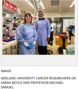
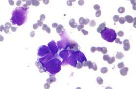
A recent study conducted by scientists working in Adelaide University and published in the journal Science Advances has shown the reason as to why certain cancers may grow and survive the body, whereas others do not. It happens that the hard mechanical stress to which the early cancer cells undergo as they are squeezed into a narrow area, causes some of the cancer cells to grow quicker, not to grow, as would otherwise be supposed.

This squeeze worked to the favor of the early breast cancer cells as scientists discovered.

The key point that was explained by the lead researcher, Professor Michael Samuel, of the Centre of Cancer Biology at Adelaide University and the Basil Hetzel Institute is that these breast cancer cells steal a particular sensor – one that our bodies rely on to sense touch – and use it to divide quickly and aid them in making their escape off the major tumour.

The process creates an indefinitely lasting mechanical memory in the breast cancer cells and it still contributes towards aggressive behaviour even after the pressure itself has been removed, Professor Samuel said.

The tumours which are solid are exposed to a lot of physical pressure when the disease is at its early stage of development, as the cancer cells grow in tissues that are limited in space, e.g. the milk ducts of the breast. Up to this day, the mechanism by which these cancer cells detect this pressure and whether or not it impacts the progression of the disease is unknown.

We have a tendency to believe that cancer is a genetic disease, but through this work we know that there is the same importance of physical forces within the tumours as the cause of cancer as there are genetic changes that cause cancer.

The researchers discovered that cancer cells respond to pressure via a molecule named PIEZO1, which is a hole in the cell that relates the interior of a cell to the exterior environment. Upon pressure stimulation, PIEZO1 enables the movement of calcium ions into the cell and subsequent signal transduction containing the Rho-ROCK pathway – a central regulator of cell movement, shape and growth.

The team demonstrated that mechanical pressure of a short duration that is obtained through compressing cancer tissue was sufficient to cause tumour growth to increase significantly. Mechanically compressed tumours in laboratory models of breast cancer became larger and the cancer cells in them fragmented faster than control groups.

In addition to promoting growth, compression was also identified to drive cancer cells into a more aggressive, invasive, state in a process known as epithelial-mesenchymal transition. When either of the PIEZO1 or the Rho-ROCK pathway had, however, been inhibited with the help of suitable drugs, compression did not propel cancer aggressiveness, making their role in this process definite.

Co-lead author Dr Sarah Boyle mentioned that one of the most significant findings was that the cancer aggressiveness effects of compression remained even after removal of the force itself.

According to Dr Boyle, even relatively short durations of pressure can lead to a mechanical memory by altering the way the DNA is packed into the cell, by chemically modifying the histone proteins.

These changes, which are called epigenetic changes, are modifications of the interpretation of the DNA code by the cell, which enables the process of switching on some genes that promote tumour growth and aggressiveness.

This type of epigenetic mechanical memory offers a molecular basis to the long term effects of short term mechanical forces on the cell level of the behaviour of tumours.

Notably, the research established that PIEZO1 is over-expressed in human breast cancers compared to normal breast tissue, and that the level of PIEZO1 differs among the patients. The high PIEZO1 levels have been linked to low patient survival implying that the identical pressure-detecting system found in test animals would probably be applicable in human cancer.

The results indicate a little-known role of mechanical pressure in the development of cancer aggressiveness and represent the PIEZO1 -Rho-ROCK pathway as a possible new therapeutic objective that can be used as an early intervention.

According to the researchers, future therapies can restrict tumour growth and invasiveness by interfering with the sensory and response of cancer cells to mechanical pressure. The results can also be applied in diagnosing the patients who are susceptible to aggressive breast cancers due to excessively high concentrations of PIEZO1.

That work has opened up a whole new field of so-called mechanotherapy – the use of treatments that disrupt the mechanical signals that tumours are dependent on to develop and spread out, as cancers grow to be mechanically responsive diseases, said Professor Samuel.

Less psychedelic, more medical magic mushrooms

The psychoactive substance of magic mushrooms, psilocybin, is under scientific scrutiny as being useful in the treatment of neuropsychiatric disorders such as depression, anxiety, substance use disorder and some neurodegenerative diseases. It can be limited to broader therapeutic uses, however, by the hallucinogenic effects. A study on the effects of psilocin, the active compound in psilocybin, on mice published in the Journal of Medicinal Chemistry by researchers synthesized modified versions of psilocin which preserve its properties but have fewer hallucinogenic-like effects than pharmaceutical-grade psilocybin.

In line with the emerging scientific view that psychedelic and serotonergic works can be decoupled, one correspondent author of the study, Andrea Mattarei, states that their findings align with this emerging school of thought. This creates the prospect of developing new therapies that are more biologically active but less hallucinogenic, which might allow developing safer and more practicable treatment options.

Mood disorders as well as certain neurodegenerative ailments such as the Alzheimer disease entail an imbalance of the neurotransmitter molecule serotonin that aids in controlling moods and other brain processes. Psychedelics have been studied to have therapeutic effects against serotonin-signaling pathways in decades by scientists. But the hallucinations that can be used along with these drugs can cause people to fear their use even in case there is a medical advantage.

Brain Image (NIH)

Therefore, a group supervised by Sara De Martin, Mattarei and Paolo Manfredi chemically engineered 5 psilocin analogs to release gradually, slowly and possibly non-hallucinogenic into the brain. The initial test of the five compounds was conducted using human plasma samples and the laboratory parameters that replicate gastrointestinal absorption. These tests have enabled the group to determine a compound they refer to as 4e as the best prospect since it exhibited desirable stability to be absorbed and allowed a slow release of psilocin – a trait that has the potential to reduce the effects of hallucinations. Notably, 4e was also active at major serotonin receptors, and at similar levels as psilocin.

The researchers then compared the impact of the same dosage of 4e on mice with pharmaceutical quality psilocybin. The team orally gave the compounds to mice and assessed the degree to which psilocin was absorbed by the bloodstream and the brain after 48 hours. The compound had the capability of penetrating the blood-brain barrier in mice treated with 4e and had a lower yet more prolonged presence of psilocin in their brain than did their psilocybin-treated counterparts. In examining the behavior of the mice, the researchers found that the 4e-treated mice had reduced the number of head twitches, a well-established oral psychiatric effect of psychedelics in rodents, with the 4e-treated mice compared to those treated with psilocybin having far fewer head twitches. This difference in behavior seemed to be linked with the quantity and the time that psilocin was released in the brain.

According to the researchers, the results of their experiments testify to the possibility of creating stable derivatives of psilocin penetrating the brain and preserving the function of serotonin receptors without acute psychotropic effects. Their mechanism of action and complete description of their biological effects will require further research before their therapeutic capacity and safety in human beings are evaluated.

The authors admit MGGM Therapeutics, LLC. funding in partnership with NeuroArbor Therapeutics Inc. Some of the authors state that they are patent holders regarding psilocin.

What is Rett Syndrome ? New study Opens Avenue to Cure This Rare Disorder

A group of researchers at the Texas Children Neurological Research Institute (NRI), Duncan, Baylor College of Medicine, found  a possible new therapy to cure Rett syndrome has been reported in the Science Translational Medicine journal, with the early signs of success in a neurodevelopmental disorder with no cure yet.

According to the corresponding author, Dr. Huda Zoghbi, the director of the Duncan NRI, Distinguished Service Professor at Baylor, and an investigator at the Howard Hughes Medical Institute, Rett syndrome is an uncommon genetic neurodevelopmental disorder that results in a developmental regression, usually past 6 to 18 months of normal development, followed by severe motor skill impairment, speech and communication impairment and communication impairment. The disorder affects girls mostly and it occurs in 1 out of 10,000 live births.

Loss-of-function mutations in MECP2 gene cause Rett syndrome, which plays a central role in normal brain functions since it controls the amount of various genes controlling neurological functions. These mutations cause the protein to disappear or code a faulty protein which cannot perform its normal function. Certain mutated forms of MeCP2 protein are also less abundant and/or reduced in terms of DNA binding, which is a key role in the functioning of this protein.

In mouse models of Rett syndrome, it has been demonstrated that the disorder is reversible, once normal MeCP2 protein is added in the brains of those mice, the conditions are reversible. Notably, it has been demonstrated by researchers that raising the concentration of a mutant MeCP2 protein that retains a minimal amount of functioning also enhances symptoms, such as survival, motor coordination and respiratory defects in mice.

The importance of this in that approximately 65 percent of patients with Rett syndrome have partially functional MeCP2, which is either less abundant or lacking DNA binding capacity, as compared to the normal level, according to the authors. Our study, on working with mouse models and cells obtained by patients with Rett syndrome, suggests evidence of concept that an increase in the levels of mutant MeCP2 in patients with the condition would be therapeutically beneficial.

Mechanism of the MECP2 gene 

The creation of therapeutics that regulate the level of MeCP2 is not that simple. A mutation to MeCP2 that is too small leads to Rett syndrome, but an excess of MeCP2 leads to another neurological disorder, MECP2 Duplication Syndrome. The balance of the question has been so delicate that it has been difficult to come up with safe and efficient treatments.

Zoghbi said that before this research, they had already learned that the brain normally forms two versions of the MeCP2 protein E1 and E2, which differ slightly. These versions are the result of the same gene, which gets processed in one direction to form E1 and in another to form E2.

Consider a gene as a blueprint of a protein. MeCP2 has four ingredients, which are e1, e2, e3 and e4. In order to produce the MeCP2-E1 protein, cells simply mix ingredients e1, e3 and e4. In order to create MeCP2-E2, the cells mix all four ingredients, which makes ingredient e2 peculiar to this form of the protein. Both versions are produced by the brain, but E1 is prevalent.

“We also learnt that no cases of Rett syndrome individuals have been reported to have mutations on E2 protein. The condition is only caused by mutations that interfere with E1 protein,” said Tirumala. This is supported in studies on mice.

“All in all, we have known that MeCP2-E2 is a single ingredient below MeCP2-E1 in the gene and it is not so abundant as E1 and not linked to Rett syndrome and is not required to support the MeCP2 activities in the brain,” Tirumala said. “This made us hypothesize that by instructing the brain cells to omit the e2 ingredient, the production of additional MeCP2-E1 protein in patients with Rett syndrome will be promoted and will lead to a better disease outcome. We have experimental evidence in the mice and cells of patients with Rett-syndrome to support our hypothesis.”

The researchers first genetically removed the ingredient e2 of normal Mecp2 gene in mice and evaluated the outcome of the deletion on the abundance of the protein and its neurological activity. Tirumala said that the results of this approach gave 50 to 60 percent MeCP2 protein increment in normal mice.

The researchers subsequently used the same strategy on the cells that were obtained using the patients with Rett syndrome that contained MECP2 mutations that decrease the abundance and activity of the protein. They removed the ingredient e2 in this mutant MECP2 gene and evaluated the impact of this mutation on the abundance of this protein and the attributes of these cells. Tirumala said that they were excited to find that MeCP2 production improved when ingredient e2 was deleted. Notably, with the degree of mutation, these cells reappeared with part or all of their usual structure, their usual electrical functions and their capability to control the level of other genes.

Lastly, the team evaluated the therapeutic potential of such an approach. Does a blocking drug of ingredient e2 elevate MeCP2 protein?

Tirumala said the value of morpholinos was tested to stimulate MeCP2 protein production in mice. “Morpholinos are artificial molecules to be used in this instance to inhibit the production of MeCP2-E2 protein by preventing the e2 ingredient to enter the cell,” explained Tirumala. “This was interesting because our morpholinos found to increase MeCP2 protein in mice tremendously.”

The work by Zoghbi and colleagues forms the basis and offers preclinical support to a treatment method of Rett syndrome involving the enhancement of MeCP2 and offering some functional recovery, Zoghbi said. Even though morpholinos are not an option due to their toxicity, analogous ones, such as antisense oligonucleotide therapies that are already being employed to treat other diseases, might be created in Rett syndrome.

Use of Boron in Proteins to Create New Treatments to Cure Cancer: Study

A large number of the most important proteins of modern medicine and science are insoluble. These comprise a host of signalling proteins and protein hormones, and all of the receptors embedded in the cell membranes, which are directed at approximately 60 percent of the active ingredients presently utilized in medicines. When the concentration of these proteins crosses some given level, they will form clumps and become useless.

This aggregation renders synthesis of these molecules in lab impossible. Since specialised production with specialised synthesing robots always needs more than a single fragment to be conjugated into a full protein, a single poorly soluble fragment of protein is usually sufficient to inhibit production. The reason is that the current techniques employed by chemists to assemble protein fragments merely perform successfully when the fragments exist in solution and in very high concentrations.

A team of researchers, headed by Jeffrey Bode, professor at the Laboratory of Organic Chemistry at ETH Zurich has now discovered how to couple even the poorly soluble portions of proteins into functional proteins. In order to do this, they utilized special properties of a chemical compound comprising an element named boron.

The slow carbon chemistry has a concentration constraint.

The only major difference between the ETH technique and the traditional strategies is in the rate of the coupling reaction. Unlike in biochemistry, which occurs extremely fast in cells of living organisms, through enzymes, reactions such as these typically need to be carried out at unnatural concentrations within the laboratory. The reason behind this is that the slower the reaction is taking place, the greater the concentration of the reacting substances should be so that the reaction processes take place as intended.

The novel coupling technique invented by the team of Bode is approximately 1000 times faster and thus was also applicable in 1000 times lower concentrations.

Boron opens up new opportunities bio-chemistry

The ETH chemists hastened the reaction by including Boron atoms to the carbon-based molecules. These are not found in natural molecules.

In several of its properties, the metalloid boron behaves in a somewhat different way. On bonding with metals, it forms very tough and heat-resistant metal alloys. Alternatively, it is capable of bonding with the nonmetals carbon, oxygen or nitrogen in the lab to form molecules that tend to have bizarre reaction characteristics. In 2010, Akira Suzuki, a Japanese researcher and Richard Heck, an American researcher, won the Nobel Prize in Chemistry due to the development of boron-based coupling reactions to enable laboratory synthesis of natural substances.

According to Bode, “We reach an ultimate limit of reaction rate with purely carbon based systems. It is further expansion into previously untapped boron based reagents that places us in a space where even the most recalcitrant reactions that bring large biological molecules together can occur in a very brief time.”

cancer cells/photo:en.wikipedia.org

Protective acids: a rocky road

As shown by Bode and colleagues in 2012, this was the first study to demonstrate that it was possible to add an element of a hitherto unexplored chemical group to proteins fragments and do so with great speed and stability. Nevertheless, this compound was not stable with strong acids hence could not be utilized in automated synthesis.

To endure the tough environment that was applied to the sensitive boron compound in normal laboratory robots, the compound would require protection in the form of a chemical packaging, but this was easier said than done. The researchers experimented with a number of strategies in four years to little effect.

The discovery was made by mistake and eventually, the discovery occurred when a doctoral student tried an experimental method that the team had indeed thought was ineffective. The resulting protective compound binds to the boron group on three sides, therefore, being unable to be terminated in the acids in protein production.

According to Bode, such fundamental research, in which there is no assurance of success, is feasible only due to the unrestricted funds provided by the Swiss National Science Foundation and ETH.

Inorganic amino acids and cancer treatment

The ETH method implies that new peptide and protein drugs or drugs of medical interest to cure cancer that are prone to clumping, can now be produced via the usual laboratory protocols.

Moreover, special amino acids that are not natural can also be incorporated in the location of choice on the poorly soluble proteins. As an example, the chemists can functionalized these building blocks in a protein in a specific way in case they wish to attach it to an active substance on a particular location. Some of the applications of antibody-drug conjugates prepared through this method include cancer treatment procedures that do not damage normal tissues.

The way in which the method will be applied to clinical practice is not yet clear. In 2020 Bode co-founded the ETH spin-out Bright Peak Therapeutics, which applications the technologies invented in his lab to build immunotherapies to fight cancer. A therapeutic agent has already entered clinical trials and the new method based on boron may assist in increasing the size of the product pipeline of the spin-off.

New recombinant mpox strain detected in UK and India, WHO urges continued monitoring

WHO confirmed that two cases of the recombinant strain – combining genomic elements of clades Ib and IIb of the monkeypox virus (MPXV) – have been identified to date: one in the United Kingdom and one in India. Both patients had recent travel histories, and neither experienced severe illness.

No secondary cases were detected following contact tracing.

WHO has published a detailed update on the two cases and the national responses.

Recombination is a natural process that can occur when two related viruses infect the same person and exchange genetic material, producing a new variant.

According to WHO, detailed genomic analysis shows the two individuals “fell ill several weeks apart with the same recombinant strain,” suggesting that additional undetected cases may exist.

What is mpox?

Mpox is an infectious disease caused by the monkeypox virus (MPXV), part of the Orthopoxvirus genus, which also includes the virus that causes smallpox.

It spreads through close physical contact, including sexual contact, and in some cases through contaminated materials or respiratory droplets.

Symptoms typically include fever, swollen lymph nodes, and rash and/or lesions.

Click here for the WHO factsheet on mpox.

Two detected cases

The case in the United Kingdom was detected in December 2025 in a traveller returning from a country in the Asia Pacific region. Initial laboratory testing identified the virus as clade Ib, but whole genome sequencing later revealed that it contained genetic regions from both clade Ib and clade IIb strains. Repeat sequencing confirmed the findings and demonstrated that the virus “can replicate and presents potential for onward transmission.”

In India, a patient who developed symptoms in September 2025 was initially classified as infected with clade II MPXV. However, following updates to global genomic databases, the virus was reclassified as the same recombinant strain identified in the UK. The Indian case represents the earliest known detection of this strain.

“Due to the small number of cases found to date, conclusions about transmissibility or clinical characterization of mpox due to recombinant strains would be premature, and it remains essential to maintain vigilance regarding this development,” WHO said.

Clinical presentations in both cases were consistent with known mpox infections.

Risk assessment unchanged

WHO’s overall risk assessment remains unchanged: the risk is assessed as moderate for men who have sex with men with new and/or multiple partners and for sex workers or others with multiple casual sexual partners, and low for the general population without specific risk factors.

At the same time, WHO cautioned that clade differentiation PCR tests alone “may not reliably identify recombinant MPXV strains,” meaning genomic sequencing is essential for detection.

“All countries should remain alert to the possibility of MPXV genetic recombination,” WHO said, urging continued epidemiological surveillance, sequencing, vaccination of at-risk groups, and infection prevention and control measures.

WHO advised that no travel or trade restrictions are warranted based on current information.

US withdrawal from WHO ‘risks global safety’, agency says in detailed rebuttal

WHO Rejects US Criticism, Defends Pandemic Response And Calls For Continued Cooperation

The World Health Organization has strongly rejected criticism from the United States administration, defending its handling of the COVID-19 pandemic and reaffirming its commitment to global cooperation on public health.

In a statement released on Saturday, the UN health agency responded to accusations from the US government that it had compromised its independence and pursued policies hostile to American interests. The organization said such claims were unfounded and reiterated that it has consistently worked with member states in good faith.

The agency also expressed hope that the United States would remain engaged in global health efforts despite recent tensions.

WHO Rejects Allegations Of Bias

Responding to accusations that it had “trashed and tarnished” the United States, the WHO said the opposite was true.

According to the statement, the organization has always sought to maintain constructive engagement with the US government while respecting its sovereignty.

It also rejected claims that the agency had followed a politicized agenda influenced by countries hostile to US interests.

“The World Health Organization has always been and remains impartial,” the statement said, adding that the organization exists to serve all countries equally and without political bias.

Defence Of COVID-19 Pandemic Response

A large portion of the statement focused on defending the WHO’s actions during the COVID-19 pandemic.

The US administration had accused the organization of obstructing the timely sharing of information and concealing failures in its response to the outbreak.

The WHO said those allegations were incorrect, arguing that it shared available information rapidly and transparently throughout the crisis while providing guidance based on the best scientific evidence at the time.

The agency clarified that while it recommended protective measures such as mask use, vaccination and physical distancing, it did not mandate lockdowns or vaccine requirements.

Early Warnings Issued In Initial Stages

According to the WHO, it began monitoring the emerging outbreak immediately after receiving reports of a cluster of pneumonia cases of unknown cause in Wuhan, China, on 31 December 2019.

The organization said it quickly contacted Chinese authorities for additional information and activated its emergency incident management system.

By the time the first death linked to the virus was reported on 11 January 2020, the WHO said it had already alerted the international community through formal channels, public statements and social media.

It also convened global health experts and issued guidance to help governments prepare their healthcare systems and protect their populations.

When the WHO Director-General declared COVID-19 a Public Health Emergency of International Concern on 30 January 2020, fewer than 100 cases had been recorded outside China and no deaths had been reported beyond the country.

During the early months of the crisis, the Director-General repeatedly warned countries that urgent action was needed to contain the virus, cautioning that the “window of opportunity is closing.”

Steps Taken To Strengthen Pandemic Preparedness

The WHO noted that several independent reviews have examined the global response to the pandemic, including assessments of the organization’s own performance.

Following these evaluations, the agency said it has introduced reforms to strengthen its ability to respond to future health emergencies.

These efforts include initiatives to improve international coordination, strengthen surveillance systems and support countries in building stronger public health infrastructure.

According to the WHO, the global systems it developed before, during and after the pandemic have helped improve preparedness worldwide.

Agency Leaves Door Open For US Engagement

Despite the current dispute, the WHO emphasised that it remains committed to working with all countries, including the United States.

The organization highlighted the recent adoption of the WHO Pandemic Agreement, which it described as a major international legal framework aimed at preventing and responding to future global health emergencies.

The agency also acknowledged the significant role the United States has historically played in advancing global public health.

As a founding member of the WHO, the US has contributed to several major achievements, including the eradication of smallpox and progress in combating diseases such as polio, HIV, tuberculosis, malaria and Ebola.

“WHO remains steadfastly committed to working with all countries in pursuit of its core mission,” the statement concluded, reaffirming its goal of ensuring the highest attainable standard of health as a fundamental right for people worldwide.

Global alliance meets in Doha to confront hunger crisis

Addressing heads of state, ministers and international partners, President of the UN General Assembly Annalena Baerbock said today’s hunger crisis is not the result of scarcity, but of inequality, conflict and policy choices.

Last year, more than 670 million people experienced hunger, and 2.3 billion faced moderate or severe food insecurity. “That is billions wondering where their next meal will come from. Parents having to see their children go to bed hungry,” she said.

This is occurring in a world that wastes over one billion meals every day.

“The crisis of hunger is not lack of food. It is entirely preventable,” she stressed, pointing to failures in access, affordability and social protection.

The meeting took place as Doha hosts the Second World Summit for Social Development, where nearly 14,000 attendees are discussing how to strengthen social systems, expand opportunity and reduce inequality.

As the planet heats, hunger spreads

Ms. Baerbock highlighted climate change as a rapidly accelerating driver of hunger. Recalling a recent visit to the Sahel, she described fertile land turned to dust as heat rises and rains fail. “This is the new frontline of food insecurity,” she said.

If global warming continues unchecked, as many as 1.8 billion additional people could face food insecurity, she warned. But limiting warming to 1.5°C, backed by investment in adaptation and resilience, could prevent millions from falling deeper into poverty.

Launched under Brazil’s G20 Presidency in 2024, the Global Alliance Against Hunger and Poverty now includes nearly 200 members – over 100 countries, regional organizations, international agencies and civil society groups. Monday’s meeting was its first at leaders’ level, aimed at accelerating practical cooperation, from scaling up social protection to strengthening climate-resilient agriculture.

“In a world of plenty – where there should be more than enough to go around – ensuring that everyone, everywhere has enough to eat is entirely possible,” Ms. Baerbock said. “A world free from hunger and poverty is not a distant aspiration. It is within reach, if we reach for it together.”


80 million more children benefiting from school meals, WFP reports

The number of children receiving school meals through government-led programmes has gone up by 20 per cent since 2020, found the latest edition of the WFP’s flagship biennial report The State of School Feeding Worldwide.

Now, nearly 80 million more children are able to enjoy nutritious meals at school, bringing the global total to approximately 466 million.

Beyond health and diet, national programmes can benefit employment, agriculture, and other sectors.

“School meals are so much more than just a plate of nutritious food – important as that is. For the vulnerable children who receive them, they are a pathway out of poverty and into a new world of learning and opportunity,” said Ms. McCain.

“They are proven to be one of the smartest, most cost-effective investments any nation can make to improve the long-term health, education and economic prosperity of future generations,” she added.

Example of what’s possible

The increase in the number of children receiving school meals comes thanks to the expansion of these programmes internationally, and especially by countries that are part of the School Meals Coalition, a network led by over 100 governments with the WFP as its secretariat.

Global funding for school meals has more than doubled, rising from $43 billion in 2020 to $84 billion last year. Africa is leading the surge with an additional 20 million children in the continent now fed through national programmes but domestic funding still remains lower in low-income countries.

“The surge in nationally funded school meal programmes is a powerful sign of what’s possible, even in challenging times. But in low-income countries, where needs are greatest, progress remains at risk as global aid shifts and domestic resources fall short,” said Carmen Burbano, director of school meals at WFP.

Meals improve learning outcomes

Children who are hungry might not attend school or could struggle to focus even if they do, according to the report. Providing meals at school can both incentivize attendance and help students stay engaged and more easily absorb educational material.

The report found that school meals are a significantly more effective way to improve the quality of education compared to other popular programmes and policies like teacher training and tech inputs.

A nutritious diet has also been associated with an increased attention span, higher cognitive function and better attendance.

“It’s only now that we’re really recognizing that the wellbeing of school children and adolescents is key to their learning ability,” said Professor Donald Bundy, co-editorial lead for the report, at a press briefing on Wednesday.

A catalyst for the economy

The report estimates that delivering school meals to 466 million children generates around 7.4 million cooking jobs globally, with further employment across logistics, farming, and supply chains.

On a national level, school meal programmes typically generate approximately 1,500 jobs for every 100,000 children.

Preliminary findings in some African countries suggest that the programmes are cost-beneficial in terms of the gains obtained in the education, health and nutrition sectors. In Malawi, for example, every $1 invested brought economic benefits ranging to $2 to $18 depending on the district.

Local procurement of school food can also create reliable and predictable markets for smallholders and family farmers, which ultimately encourages crop diversification, boosts rural economies, and fosters sustainable agricultural practices.

‘Shift the narrative’ on suicide to prevent loss of 720,000 lives annually

Speaking on World Suicide Prevention Day, which is marked annually on 10 September, WHO’s Tedros Adhanom Ghebreyesus said that “each life lost leaves a profound impact on families, friends, colleagues and entire communities.”

All age groups are affected by suicide and was the third leading cause of death among 15–29-year-olds globally in 2021, the last year for which data has been gathered by WHO.

Suicide does not just occur in high-income countries and impacts all regions of the world.

Close to three quarters of global suicides occurred in low and middle-income countries in 2021.

The average number of suicides across the world in 2021 was 8.9 per 100,000 people.

In Africa the figure stood at 11.5, while in both Europe and Southeast Asia the number of suicides was recorded at 10.1 per 100,000 people.

Globally, the lowest suicide rate was in the Eastern Mediterranean region at 4.0 per 100,000, while in the Western Pacific it was 7.5 per 100,000.

Who’s at risk?

The link between suicide and mental disorders, in particular, depression and alcohol use disorders, and a previous suicide attempt is well established in high-income countries.

However, many suicides happen impulsively in moments of crisis with a breakdown in the ability to deal with life stresses, such as financial problems, relationship disputes, or chronic pain and illness.

In addition, experiencing conflict, disaster, violence, abuse or loss and a sense of isolation are strongly associated with suicidal behaviour.

Suicide rates are also high among vulnerable groups who experience discrimination, such as refugees and migrants, indigenous peoples, lesbian, gay, bisexual, transgender, intersex (LGBTI) persons and incarcerated prisoners.

Moving from silence to openness

We must move from silence to openness, from stigma to empathy, and from neglect to support,” said Dr. Tedros.

“We must create environments where people feel safe to speak up and seek help,” he said.

“Shifting the narrative on suicide also means driving systemic change, where governments prioritise and invest in quality mental health care and policies to ensure everyone gets the support they need.”

According to the 2024 Mental Health Atlas report by WHO, median government spending on mental health has remained at a modest 2 percent of total health budgets since 2017.

Moreover, there is a significant disparity between high-income and low-income nations. Whilst high-income nations allocate up to $65 per person to mental health, low-income nations spend as little as $0.04.

WHO recognizes mental health as a universal human right.

Effective prevention measures

WHO says that there are effective measures that can be taken to prevent suicide and self-harm.

LIVE LIFE, the agency’s initiative for suicide prevention, recommends the following key effective evidence-based interventions:

  • limit access to the means of suicide (eg, pesticides, firearms, certain medications);
  • interact with the media for responsible reporting of suicide;
  • foster socio-emotional life skills in adolescents;
  • early identify, assess, manage and follow up anyone who is affected by suicidal behaviours.

Child obesity level surpasses underweight cases worldwide for the first time, UNICEF warns

One in 10 children aged 5 to 19 – 188 million worldwide – are now living with obesity, placing them at heightened risk of chronic diseases such as type-2 diabetes, heart conditions, and certain cancers.

“When we talk about malnutrition, we are no longer just talking about underweight children,” said UNICEF Executive Director Catherine Russell.

“Obesity is a growing concern that can impact the health and development of children. Ultra-processed food is increasingly replacing fruits, vegetables and protein at a time when nutrition plays a critical role in children’s growth, cognitive development and mental health”, she added.

The report, Feeding Profit: How Food Environments are Failing Children, draws on data from over 190 countries and highlights a stark shift.

One in five overweight

Since 2000, the number underweight among five to 19-year-olds has dropped from nearly 13 per cent to 9.2 per cent.

In the same period, obesity has tripled, from three per cent to 9.4 per cent. Today, obesity rates exceed underweight in every region except sub-Saharan Africa and South Asia.

The situation is particularly acute in the Pacific Islands, where traditional diets have been displaced by cheap, energy-dense imported foods.

High-income countries are not exempt: 27 per cent of children in Chile, and 21 per cent in both the United States and United Arab Emirates, are affected.

Globally, one in five children and adolescents, or 391 million, are overweight, with nearly half now classified as obese.

Children are considered overweight when they are significantly heavier than what is healthy for their age, sex and height.

Obesity is a severe form of overweight and leads to a higher risk of developing insulin resistance and high blood pressure, as well as life-threatening diseases later in life, including type-2 diabetes, cardiovascular disease, and certain cancers.

A consumer in Mongolia eats a sugary desert.

Marketing to blame

The report points to powerful commercial forces shaping these outcomes. Ultra-processed and fast foods, high in sugar, salt, unhealthy fats and additives, dominate children’s diets and are aggressively marketed, influencing children’s diets.

In a UNICEF poll of 64,000 young people across 170 countries, 75 per cent reported seeing ads for sugary drinks, snacks, or fast food in the previous week.

Sixty per cent said the ads made them want to eat the products. Even in conflict-affected countries, 68 per cent of young people said they were exposed to these advertisements.

These patterns, UNICEF warns, carry staggering economic consequences. By 2035, the global cost of overweight and obesity levels is projected to exceed $4 trillion annually. In Peru alone, obesity-related health issues could cost over $210 billion across a generation.

Government must act

Still, some governments are taking action. Mexico – where sugary drinks and ultra-processed foods make up 40 per cent of children’s daily calories – has banned their sale in public schools, improving food environments for more than 34 million children.

UNICEF is urging governments worldwide to follow suit with sweeping reforms: mandatory food labelling, marketing restrictions, and taxes on unhealthy products; bans on junk food in schools; stronger social protection programmes; and safeguards to shield policymaking from industry interference.

“In many countries we are seeing the double burden of malnutrition, the existence of stunting and obesity. This requires targeted interventions,” said Ms. Russell.

Nutritious and affordable food must be available to every child to support their growth and development. We urgently need policies that support parents and caretakers to access nutritious and healthy foods for their children”, she concluded.
 

WHO sounds alarm as mental health conditions soar past one billion worldwide

Disorders such as anxiety and depression are exacting a heavy toll on individuals, families and economies, yet most countries are failing to provide adequate support.

Mental health problems are widespread across every society and age group and remain the second leading cause of long-term disability. They drive up healthcare costs for families and governments while costing the global economy an estimated $1 trillion each year in lost productivity, UN health experts said.

Way off track

The findings are detailed in two new reports: World mental health today and the Mental Health Atlas 2024.

Together, they show that while there has been some progress since 2020, the world is still far off track in tackling the scale of the crisis. The reports will help to inform debate at a UN high-level meeting on noncommunicable diseases and mental health, to be held late this month in New York.

Transforming mental health services is one of the most pressing public health challenges,” said WHO Director-General Dr Tedros Adhanom Ghebreyesus.

Investing in mental health means investing in people, communities and economies, an investment no country can afford to neglect. Every leader has a responsibility to act urgently and to ensure mental health care is treated not as a privilege, but as a basic right.”

Troubling gaps, uneven progress

The reports highlight several stark findings:

  • Women are disproportionately affected by mental health conditions, with anxiety and depression most common among both sexes.
  • Suicide claimed an estimated 727,000 lives in 2021 and is a leading cause of death among young people. On current trends, the world will fall far short of the UN target to reduce suicide deaths by a third by 2030, managing only a 12 per cent reduction.
  • Median government spending on mental health remains at just two per cent of health budgets, unchanged since 2017. While high-income countries spend up to $65 per person on mental health, low-income countries spend as little as four cents.
  • The mental health workforce is dangerously thin in many regions. There are just 13 mental health workers for every 100,000 people worldwide.
  • Fewer than one in 10 countries has fully moved to community-based care, with most still relying heavily on psychiatric hospitals. Almost half of inpatient admissions are involuntary, and more than one in five patients remain hospitalised for over a year.

Despite these challenges, there have been some positive developments. More countries are integrating mental health into primary healthcare and expanding early intervention programmes in schools and communities.

Over 80 per cent of countries now include mental health and psychosocial support in emergency response, up from less than 40 per cent in 2020. Telehealth services are also becoming more widely available, though access is still uneven.

Call for systemic change

WHO is urging governments to step up investment and reform, warning that the current pace of progress is too slow to meet global goals. Key priorities include:

  • Fairer financing of mental health services
  • Stronger legal protection and rights-based legislation
  • Greater investment in the mental health workforce
  • Accelerated shift towards community-based, person-centred care

The UN health agency stresses that mental health should be treated as a fundamental human right. Without urgent action, millions will continue to suffer without support, and societies will bear rising social and economic costs.

For more information on how the UN overall is advocating for more resources to support mental health and wellbeing, check out this story from our colleagues at www.un.org