- Many breast cancer therapies work by causing DNA replication stress, but triple-negative breast cancer (TNBC) cells survive this stress, even at high levels
- TNBC cells overexpress an enzyme, RNase H2, that helps them survive the DNA damage caused by replication stress
- In this preclinical study, blocking RNase H2 directly damaged cancer cells and also activated the immune system, making this a promising therapeutic approach.
Blocking RNase H2 Enzyme Shows Promise Against Aggressive Breast Cancer
Scientists at University of Texas MD Anderson Cancer Center have identified a crucial enzyme that helps one of the most difficult forms of breast cancer withstand treatment, opening up a potential new therapeutic pathway.
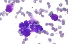
The study, published in Cell Reports Medicine, focuses on RNase H2—an enzyme that appears to enable triple-negative breast cancer (TNBC) cells to survive intense DNA damage caused during treatment. TNBC is widely considered one of the most aggressive breast cancer subtypes due to its resistance to many standard therapies.
Led by researcher Shiaw-Yih Lin, the team found that RNase H2 plays a dual role. While it helps cancer cells cope with DNA replication stress, blocking the enzyme both damages tumour DNA and triggers the body’s immune system, effectively turning the cancer’s survival mechanism against itself.
Lin described this as a “one-two punch,” where inhibiting RNase H2 not only disrupts the tumour’s ability to manage stress but also activates immune signals that draw T cells to attack the cancer.
Why replication stress matters
Replication stress occurs when cells struggle to copy their DNA accurately, leading to structural damage. Many cancer treatments deliberately induce this stress to kill tumour cells. However, TNBC cells have developed ways to tolerate and survive it, allowing the disease to persist and progress.
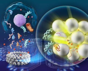
One key contributor to this stress is the build-up of RNA fragments within DNA. RNase H2 typically removes these fragments to maintain genomic stability.
The enzyme’s hidden role in cancer survival
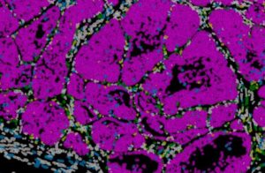
The researchers discovered that RNase H2 is significantly overproduced in TNBC tumours and is linked to poorer patient outcomes. This suggests that the enzyme helps cancer cells adapt to and survive high levels of DNA damage.
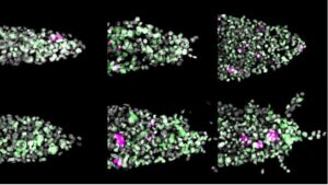
When the team blocked RNase H2—either through genetic methods or experimental drugs—tumour cells experienced heightened replication stress, leading to reduced tumour growth in preclinical models. At the same time, the resulting DNA damage activated the innate immune system, which then signalled T cells to target the cancer.
Implications for future treatment
Although the findings are still at a preclinical stage, they point to RNase H2 as a promising drug target. Inhibitors of the enzyme are already under development, and the study suggests they could be used alongside existing therapies.
Notably, blocking RNase H2 was found to enhance the effectiveness of ATR and PARP inhibitors—two classes of drugs already used in cancer treatment—raising the possibility of combination therapies in future clinical trials.
If validated in humans, this approach could offer a more effective strategy against TNBC, a cancer subtype that has long posed challenges due to its limited treatment options.
Also Read:
How being squeezed contributes to risk of breast cancer cells
Study Links High-Fat Diets To More Aggressive Breast Cancer Behavior