Researchers find potential one-two punch against triple-negative breast cancer

  • Many breast cancer therapies work by causing DNA replication stress, but triple-negative breast cancer (TNBC) cells survive this stress, even at high levels
  • TNBC cells overexpress an enzyme, RNase H2, that helps them survive the DNA damage caused by replication stress
  • In this preclinical study, blocking RNase H2 directly damaged cancer cells and also activated the immune system, making this a promising therapeutic approach.

Blocking RNase H2 Enzyme Shows Promise Against Aggressive Breast Cancer

Scientists at University of Texas MD Anderson Cancer Center have identified a crucial enzyme that helps one of the most difficult forms of breast cancer withstand treatment, opening up a potential new therapeutic pathway.

The study, published in Cell Reports Medicine, focuses on RNase H2—an enzyme that appears to enable triple-negative breast cancer (TNBC) cells to survive intense DNA damage caused during treatment. TNBC is widely considered one of the most aggressive breast cancer subtypes due to its resistance to many standard therapies.

Led by researcher Shiaw-Yih Lin, the team found that RNase H2 plays a dual role. While it helps cancer cells cope with DNA replication stress, blocking the enzyme both damages tumour DNA and triggers the body’s immune system, effectively turning the cancer’s survival mechanism against itself.

Lin described this as a “one-two punch,” where inhibiting RNase H2 not only disrupts the tumour’s ability to manage stress but also activates immune signals that draw T cells to attack the cancer.

Why replication stress matters

Replication stress occurs when cells struggle to copy their DNA accurately, leading to structural damage. Many cancer treatments deliberately induce this stress to kill tumour cells. However, TNBC cells have developed ways to tolerate and survive it, allowing the disease to persist and progress.

One key contributor to this stress is the build-up of RNA fragments within DNA. RNase H2 typically removes these fragments to maintain genomic stability.

The enzyme’s hidden role in cancer survival

The researchers discovered that RNase H2 is significantly overproduced in TNBC tumours and is linked to poorer patient outcomes. This suggests that the enzyme helps cancer cells adapt to and survive high levels of DNA damage.

When the team blocked RNase H2—either through genetic methods or experimental drugs—tumour cells experienced heightened replication stress, leading to reduced tumour growth in preclinical models. At the same time, the resulting DNA damage activated the innate immune system, which then signalled T cells to target the cancer.

Implications for future treatment

Although the findings are still at a preclinical stage, they point to RNase H2 as a promising drug target. Inhibitors of the enzyme are already under development, and the study suggests they could be used alongside existing therapies.

Notably, blocking RNase H2 was found to enhance the effectiveness of ATR and PARP inhibitors—two classes of drugs already used in cancer treatment—raising the possibility of combination therapies in future clinical trials.

If validated in humans, this approach could offer a more effective strategy against TNBC, a cancer subtype that has long posed challenges due to its limited treatment options.

Also Read:

How being squeezed contributes to risk of breast cancer cells

Study Links High-Fat Diets To More Aggressive Breast Cancer Behavior

The battle of the sexes in the egg

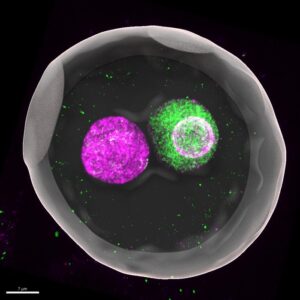
The sperm and the egg cell’s nuclei compete for size directly after fertilization and this is necessary for the proper embryonic development. The mouse study with Kobe University participation finally gives meaning to a phenomenon biologists have known for decades.

At the beginning of a new individual’s life, an egg and a sperm fuse to form what biologists call a “zygote,” a fertilized egg. In mammals such as mice and humans, the DNA-carrying nuclei of these cells don’t fuse right away but remain separate as so-called “pronuclei” until just before the first cell division. “This fact has been known for decades, but no one really understood why this separation exists. People just treated it as a descriptive feature rather than something to be tested experimentally,” says Kobe University developmental biologist KYOGOKU Hirohisa. If anything, separate nuclei may increase the risk of errors during the first cell division. In assisted reproduction, embryos in which the maternal and paternal pronuclei are fused early have therefore attracted interest, but these embryos are known to have a lower likelihood of developing to term.

Kyogoku specializes in studying early development directly after fertilization. “The DNA an individual inherits from their mother and father differs not only in its genetic information. It is also known that the maternal genome has a lot of chemical modifications, like little tags, on the molecules around which the DNA is wrapped. The genome from the sperm, on the other hand, has virtually no such modifications. But these proper modifications are essential for embryonic development,” he explains. Thus suspecting that the fusion of pronuclei might have an influence on gene regulation, he teamed up with researchers from RIKEN and, using his special cell manipulation techniques, took a close look at what happened to these modifications when the pronuclei are fused and under other conditions.

In the journal Nature, the team now published that they found that the size of the pronuclei is essential for maintaining the regulatory modifications. Whenever the size of the nucleus was large, either through manipulation or because of the premature fusing of the maternal and paternal pronuclei, the degree of the chemical tagging became low. And when they investigated what keeps the pronuclei’s sizes small in the natural, separated state, they found that the two pronuclei engage in a race to absorb factors that regulate nucleus growth. This then yielded a clear mechanistic explanation for why the developmental potential of fused nuclei is smaller: Because the single nucleus doesn’t have to compete for these factors it ends up much larger and as a result the genome’s regulatory tags get lost. “I find it exciting that a very familiar structure — two separate nuclei in a fertilized egg — turns out to have a clear and functional role, that is, it actively creates a competitive environment inside the cell which helps maintain proper regulation and supports development,” says Kyogoku.

Caption
In mammals such as mice and humans, the DNA-carrying nuclei of these cells don’t fuse right away but remain separate as so-called “pronuclei” until just before the first cell division. This 3D-reconstructed image shows a mouse zygote (fertilized egg), where maternal (magenta) and paternal (green) genomes are enclosed in separate pronuclei. “This fact has been known for decades, but no one really understood why this separation exists,” says Kobe University developmental biologist KYOGOKU Hirohisa. Credit:  KYOGOKU Hirohisa

To test their theory, the team temporarily introduced into zygotes with prematurely fused pronuclei an additional pronucleus to re-establish competition. And indeed, this intervention successfully limited nuclear size and partially restored the regulatory marks as well as the developmental potential.

The Kobe University work opens a whole new chapter of research into the very first steps a new life takes. “Even at the beginning of life, spatial organization is not just incidental but fundamentally important,” explains Kobe University biologist Kyogoku. He continues, “This result brings us one step closer to understanding the physical and biological principles that explain why early embryos are both robust and error-prone at the same time.”


Also Read:

Scientists Develop Smart Oxide That Stores Power And Shows Charge Status

Primary Breast Tumours Found To Already Contain Cells With Metastatic Potential

Exclusive Breastfeeding Linked To Lasting DNA Changes, Study Finds

Babies who are exclusively breastfed for at least three months show distinct biological markers in their blood compared to those who are not breastfed, according to a major international study.


DNA Gaps: Why Most Neanderthal Men Preferred to Sleep With Modern Female Humans?

The latest genetic study indicates that initial interactions between Neanderthals and modern humans were uneven, with some indication that most of the Neanderthals were men who slept with female modern humans, which could be the reason behind long term gaps in human DNA.

The experiment conducted by researcher, Alexander Platt and other researchers, investigates the distribution of Neanderthal genetic material in current human beings. Although the majority of those not in Africa have some traces of Neanderthal ancestry, these have been distributed unevenly throughout the human genome.

A particularly interesting characteristic is the existence of so-called Neanderthal deserts – large areas of the DNA, in which the genetic material of Neanderthals is virtually nonexistent. These deletions are more pronounced in the X chromosome and this poses a question on how the ancient interbreeding process occurred.

There has been long speculation among scientists as to whether these deletions were due to natural selection (whereby the deleterious Neanderthal genes are becoming more and more extinct) or that the interbreeding itself is the cause.

The researchers reversed the question to investigate. They did not simply study the Neanderthal DNA of the contemporary human beings, but rather the remnants of the early modern human DNA in the Neanderthal genomes. The comparison of these with genetic data of the sub-Saharan African populations, most of which do not have Neanderthal ancestry, helped the team recreate ancient gene flow patterns between the two groups.

Great Imbalances in DNA

Their results showed a great imbalance: the proportion of the modern human DNA in the Neanderthal X chromosomes was much higher than anticipated- approximately 62 percent higher. Researchers believe that such an asymmetry can best be attributed to the possibility that the vast majority of the interbreeding took place between male Neanderthals and female modern humans.

This would limit the survival of Neanderthal X-linked DNA into the subsequent generations of human population because males can only transmit their X chromosome to the females. This would over time lead to the low concentration of Neanderthal genetic material on the human X chromosome today.

The paper also indicates that social or behavioural influences, including mate preferences, could have contributed to the development of such patterns, but demographic influence, such as the variation in number or migration cannot be disqualified.

Natural Selection Behind Imbalance?

Moreover, this imbalance was probably supported by natural selection. Dangerous or incompatible genes of Neanderthals especially those associated with significant biological functions might have been gradually eliminated in human gene pool across generations.

The results provide a new understanding of the complicated relationships between the early human groups and the ones closest to their evolutionary lineage not only regarding genetic inheritance but also on social process that might have influenced the evolution of humans.

Using the combination of genomic evidence and evolutionary modelling, researchers indicate that the study is leading scientists nearer to the realization of how ancient interbreeding events still impact the genetic landscape of modern humans.

Also Read:

Neanderthal gene in modern women helps give birth to more children, says study

Research establishes traces of Neandertal DNA present in genome of modern humans


Research establishes traces of Neandertal DNA present in genome of modern humans

“Ancient DNA caused a revolution in how we think about human evolution,” says Steven Churchill, co-author of the study and a professor of evolutionary anthropology at Duke University. “We often think of evolution as branches on a tree, and researchers have spent a lot of time trying to trace back the path that led to us, Homo sapiens. But we’re now beginning to understand that it isn’t a tree – it’s more like a series of streams that converge and diverge at multiple points.”

“Our work here gives us a deeper understanding of where those streams came together,” says Ann Ross, corresponding author of the study and a professor of biological sciences at North Carolina State University.

Neanderthal/Photo:en.wikipedia.org

“The picture is really complicated,” Churchill says. “We know there was interbreeding. Modern Asian populations seem to have more Neandertal DNA than modern European populations, which is weird – because Neandertals lived in what is now Europe. That has suggested that Neandertals interbred with what are now modern humans as our prehistoric ancestors left Africa, but before spreading to Asia. Our goal with this study was to see what additional light we could shed on this by assessing the facial structure of prehistoric humans and Neandertals.”

“By evaluating facial morphology, we can trace how populations moved and interacted over time,” Ross explains. “And the evidence shows us that the Near East was an important crossroads, both geographically and in the context of human evolution.”

For this study, the researchers collected data on craniofacial morphology from the published literature. This ultimately resulted in a data set including 13 Neandertals, 233 prehistoric Homo sapiens, and 83 modern humans.

The researchers focused on standard craniofacial measurements, which are reproducible, and used those measurements to assess the size and shape of key facial structures. This then allowed the researchers to do an in-depth analysis to determine whether a given human population was likely to have interbred with Neandertal populations, as well as the extent of that likely interbreeding.

“Neandertals had big faces,” Churchill says. “But size alone doesn’t establish any genetic link between a human population and Neandertal populations. Our work here involved a more robust analysis of the facial structures.”

The researchers also accounted for environmental variables that are associated with changes in human facial characteristics, to determine the likelihood that connections they established between Neandertal and human populations were the result of interbreeding rather than other factors.

“We found that the facial characteristics we focused on were not strongly influenced by climate, which made it easier to identify likely genetic influences,” Ross says. “We also found that facial shape was a more useful variable for tracking the influence of Neandertal interbreeding in human populations over time. Neandertals were just bigger than humans. Over time, the size of human faces became smaller, generations after they had bred with Neandertals. But the actual shape of some facial features retained evidence of interbreeding with Neandertals.”

“This was an exploratory study,” Churchill says. “And, honestly, I wasn’t sure this approach would actually work – we have a relatively small sample size, and we didn’t have as much data on facial structures as we would have liked. But, ultimately, the results we got are really compelling.

“To build on this, we’d like to incorporate measurements from more human populations, such as the Natufians, who lived more than 11,000 years ago on the Mediterranean in what is now Israel, Jordan and Syria.”

Making mosquitoes self-destruct

Researchers at the University of California, Riverside have developed transgenic mosquitoes that stably express the Cas9 enzyme in their germline. The addition of Cas9 will enable the use of the CRISPR gene editing tool to make efficient, targeted changes to the mosquitoes’ DNA.

As proof of concept, the researchers used the system to disrupt cuticle, wing, and eye development, producing completely yellow, three-eyed and wingless mosquitoes. Their long-term goal is to use Cas9-expressing mosquitoes together with another technology — called gene drives — to insert and spread genes that suppress the insects while avoiding the resistance that evolution would typically favor. Aedes aegypti are major carriers of dengue, chikungunya, yellow fever, and zika viruses, and are rapidly becoming resistant to commonly used pesticides.

Published today in the Proceedings of the National Academy of Sciences (PNAS), the study was led by Omar Akbari, an assistant professor of entomology in UCR’s College of Natural and Agricultural Sciences and a member of the university’s Institute for Integrative Genome Biology.

Previous efforts to use genome editing to prevent mosquitoes from spreading pathogens have been hampered by low mutation rates, poor survival of edited mosquitoes, and inefficient transmission of disrupted genes to offspring. Akbari and colleagues developed transgenic mosquitoes that stably express a bacterial Cas9 enzyme in the germline, enabling highly efficient genome editing using the CRISPR system. CRISPR works like a pair of molecular scissors, cutting out and replacing specific DNA sequences based on a ribonucleic acid (RNA) guide. In the paper, the team used the system to disrupt genes that control vision, flight and feeding, resulting in mosquitoes with an extra eye, malformed wings, and defects in eye and cuticle color, among other changes.

Akbari said these strains represent the first step toward using gene drive systems to control mosquito populations and reduce the diseases they spread.

“These Cas9 strains can be used to develop split-gene drives which are a form of gene-drive by which the Cas9 and the guide RNA’s are inserted at separate genomic loci and depend on each other for spread. This is the safest way to develop and test gene drives in the laboratory to ensure no spread into the wild,” Akbari said.

Gene drives greatly increase the odds that a gene or set of genes will be passed on to offspring — from 50 percent to 99 percent. This number can potentially increase to 100 percent when a target gene is disrupted in multiple sites, a technique called multiplexing that has recently been mathematically modeled by Akbari and colleagues at UC Berkley.

Gene drives can be used to bias genetic inheritance in favor of rapidly spreading, self-destructive genes — such as those that disrupt fertility — and could be an environmentally friendly and cost-effective way to suppress populations of disease-spreading insects.

“Next steps should be undertaken to identify the regulatory sequences that can be used to express the guide RNAs from the genome, and once these sequences are identified developing gene drives in the species should be turnkey,” Akbari said.

New blood test may transform the way cancer is monitored and treated

Stanford University scientists have described a new type of test that can detect genetic mutations in minute amounts of DNA released from cancer cells into the blood. The test, which is called single color digital PCR, requires only a fraction of a tube of blood and can detect as few as three mutation-bearing molecules in a single reaction. According to the report in The Journal of Molecular Diagnostics, this highly sensitive test has the potential to be personalized to recognize mutations unique to any individual cancer.

“For monitoring patient tumors, only a handful of blood tests are available which are limited to only several types of cancers. Nearly all cancer patients require monitoring by whole body imaging, which can be costly, complex, and time-consuming. In contrast, molecular tests like the one we have developed will enable patients to be monitored at every visit, and thus have the potential for quickly tracking cancer growth and spread. Moreover, the test’s rapid turnaround and relatively low cost, especially compared to next-generation DNA sequencing, provide a potential opportunity for universal monitoring of more patients than is currently done,” explained lead investigator Hanlee P. Ji, MD, Associate Professor in the Department of Medicine at Stanford University and Senior Associate Director of the Stanford Genome Technology Center.

The report describes the use of the test to analyze samples from six patients. Five patients were previously diagnosed with colorectal cancer and one with cholangiocarcinoma.

After generation of customized mutation detection assays, the researchers were able to identify tumor-derived circulating DNA from three out of six patients. In one patient, the assay was able to show the presence of three different mutations. The three patients, whose samples did not show elevated cancer DNA, were undergoing active treatment at the time of collection.

The single-color digital PCR test offers several advantages over other methods of circulating tumor DNA analysis, compared to next-generation targeted sequencing and fluorescent probe-based digital PCR assays. The main advantage is that the new technique does not rely on pre-amplification, which can introduce errors and biases.

“This test is simple enough to set up and analyze without extensive training, and therefore, it can be implemented by anyone, making it highly accessible to any laboratory. It has been truly motivating to work with a technology that will help transform the way that we monitor and treat individuals with cancer. I am excited to share our findings with the cancer research community,” noted lead author and researcher Christina Wood Bouwens, of the Stanford Genome Technology Center and the Division of Oncology, Department of Medicine, Stanford University School of Medicine, Stanford, California.

Attitudes on human genome editing vary, but reach consensus on holding talks

An international team of scientists announced they had successfully edited the DNA of human embryos. As people process the political, moral and regulatory issues of the technology — which nudges us closer to nonfiction than science fiction — researchers at the University of Wisconsin-Madison and Temple University show the time is now to involve the American public in discussions about human genome editing.

In a study published Aug. 11 in the journal Science, the researchers assessed what people in the United States think about the uses of human genome editing and how their attitudes may drive public discussion. They found a public divided on its uses but united in the importance of moving conversations forward.

“There are several pathways we can go down with gene editing,” says UW-Madison’s Dietram Scheufele, lead author of the study and member of a National Academy of Sciences committee that compiled a report focused on human gene editing earlier this year. “Our study takes an exhaustive look at all of those possible pathways forward and asks where the public stands on each one of them.”

Compared to previous studies on public attitudes about the technology, the new study takes a more nuanced approach, examining public opinion about the use of gene editing for disease therapy versus for human enhancement, and about editing that becomes hereditary versus editing that does not.

The research team, which included Scheufele and Dominique Brossard — both professors of life sciences communication — along with Michael Xenos, professor of communication arts, first surveyed study participants about the use of editing to treat disease (therapy) versus for enhancement (creating so-called “designer babies”). While about two-thirds of respondents expressed at least some support for therapeutic editing, only one-third expressed support for using the technology for enhancement.

Diving even deeper, researchers looked into public attitudes about gene editing on specific cell types — somatic or germline — either for therapy or enhancement. Somatic cells are non-reproductive, so edits made in those cells do not affect future generations. Germline cells, however, are heritable, and changes made in these cells would be passed on to children.

Public support of therapeutic editing was high both in cells that would be inherited and those that would not, with 65 percent of respondents supporting therapy in germline cells and 64 percent supporting therapy in somatic cells. When considering enhancement editing, however, support depended more upon whether the changes would affect future generations. Only 26 percent of people surveyed supported enhancement editing in heritable germline cells and 39 percent supported enhancement of somatic cells that would not be passed on to children.

“A majority of people are saying that germline enhancement is where the technology crosses that invisible line and becomes unacceptable,” says Scheufele. “When it comes to therapy, the public is more open, and that may partly be reflective of how severe some of those genetically inherited diseases are. The potential treatments for those diseases are something the public at least is willing to consider.”

Beyond questions of support, researchers also wanted to understand what was driving public opinions. They found that two factors were related to respondents’ attitudes toward gene editing as well as their attitudes toward the public’s role in its emergence: the level of religious guidance in their lives, and factual knowledge about the technology.

Those with a high level of religious guidance in their daily lives had lower support for human genome editing than those with low religious guidance. Additionally, those with high knowledge of the technology were more supportive of it than those with less knowledge.

While respondents with high religious guidance and those with high knowledge differed on their support for the technology, both groups highly supported public engagement in its development and use. These results suggest broad agreement that the public should be involved in questions of political, regulatory and moral aspects of human genome editing.

“The public may be split along lines of religiosity or knowledge with regard to what they think about the technology and scientific community, but they are united in the idea that this is an issue that requires public involvement,” says Scheufele. “Our findings show very nicely that the public is ready for these discussions and that the time to have the discussions is now, before the science is fully ready and while we have time to carefully think through different options regarding how we want to move forward.”

Early puberty may mean less time in education for girls

The age at which girls have their first period may influence how long they stay in education.

The findings come from a study in which researchers have tried to untangle the effect of the age at first period from other complex factors that might affect time spent in education, revealing that young women who start their periods earlier may spend less time in the education system.

Previous research has indicated girls who reach sexual maturity earlier may be more prone to developing depression and, in low and middle income countries, more vulnerable to early pregnancy and negative sexual health outcomes, but whether it affects how long they spend in education was unclear.

Studies trying to pick apart the link between the age at which a girl has her first period — called menarche — and how long they spend in education can be muddied by numerous factors, including obesity, socio-economic status, and parental education level.

Now, a team led by researchers at Imperial College London has tried to untangle this complex relationship by turning to genetic markers as a proxy for the age of first period. Using a statistical method called Mendelian randomization, they attempted to remove the influence of external factors such as diet and lifestyle — which are known to be associated with both early menarche and less time in education.

By using genetic markers known to be associated with menarche, the researchers have revealed an impact of the age of first menstruation on the amount of time spent in education.

“It’s well established that the length of time that someone spends in education can have repercussions later on in life,” said Dr Dipender Gill, a Wellcome Trust Clinical Research Fellow at Imperial and lead author of the study. “It is associated with socio-economic status, rates of depression, risk-taking behaviour and a range of health outcomes, so clearly time spent in education is important. This study identifies that the age of puberty may have an effect on the length of time that women spend in education.”

In the study, published in the journal Behaviour Genetics, researchers looked at data from more than 180,000 European women, where 122 points in the genome where a single ‘letter’ difference in the DNA — called a single nucleotide polymorphism (SNP) — were associated with the onset of menstruation in girls.

The effects of these markers on time spent in education were then estimated using a separate dataset including more than 118,000 women over the age of 30 and of European descent, where participants had provided the number of years spent in education.

Analysis revealed a small but statistically significant causal link between markers for age at menarche and the length of time women spent in education. The findings showed that on average, starting menstruation one year later was associated with approximately an additional 53 days spent in education.

According to the researchers, one possible explanation for the observed effect could be due to young women being treated as more mature due to physical changes, while their emotional development takes time to catch up. Such a delay between physical and mental maturation may give rise to factors which lead to less time spent in education, such as increased risk-taking behaviour, or a failure to adapt psychologically to changes in how they are treated.

The group reports that the data could be skewed by women self-reporting the age they had their first period. In addition, the extended age ranges of women in the study group — born over nine decades (1901 to 1989) — overlaps with societal changes and the establishment of educational programmes. They add that the findings cannot be used to predict how long a young woman might stay in education, based on her age at her first period.

According to the researchers, now that a link has been established, the next step is to work out why age of menarche is having such an effect.

“Once we understand the mechanism, it might give us the opportunity address the discrepancies that we’re seeing,” explained Dr Gill.

“Going through puberty is associated with various physiological and psychological changes,” he adds. “It might be that girls who go through puberty earlier are less well-equipped to deal with the pressures. They may be suffering as a result and this might be manifest in the length of time that they spend in school.”

‘Age at menarche and time spent in education: a Mendelian Randomization study’ by Gill, D. et al, is published in the journal Behavior Genetics.

Scientists discover unknown virus in ‘throwaway’ DNA

A chance discovery has opened up a new method of finding unknown viruses.

In research published in the journal Virus Evolution, scientists from Oxford University’s Department of Zoology have revealed that Next-Generation Sequencing and its associated online DNA databases could be used in the field of viral discovery. They have developed algorithms that detect DNA from viruses that happen to be in fish blood or tissue samples, and could be used to identify viruses in a range of different species.

Next-Generation Sequencing has revolutionised genomics research and is currently used to study and understand genetic material. It allows scientists to gather vast amounts of data, from a single piece of DNA, which is then collated into huge, online, genome databases that are publicly accessible.

Dr Aris Katzourakis and Dr Amr Aswad, Research Associates at Oxford’s Department of Zoology, initially discovered the new use for the database, by chance. While looking for an ancient herpes virus in primates, they found evidence of two new undocumented viruses.

Spurred by their accidental discovery, they set out to see if they could intentionally achieve the same result. In a separate project to find new fish-infecting herpes viruses, they used the technique to examine more than 50 fish genomes for recognisable viral DNA. Sure enough, in addition to the herpes viruses they were expecting to find, the researchers identified a distant lineage of unusual viruses – that may even be a new viral family. The traits were found scattered in fragments of 15 different species of fish, including the Atlantic salmon and rainbow trout.

To confirm that the viral evidence was not simply a fluke, or a data processing error, they tested additional samples from a local supermarket and sushi restaurant. The same viral fragments were found in the bought samples.

Study author Dr Aris Katzourakis, from Oxford University’s Department of Zoology, said: ‘In the salmon genome we found what seems to be a complete and independent viral genome, as well as dozens of fragments of viral DNA that had integrated into the fish DNA. We know from recent studies that viruses are able to integrate into the genome of their host, sometimes remaining there for millions of years. In this case, it looks like the virus may have acquired the ability to integrate by stealing a gene from the salmon itself, which explains how it has become so widespread in the salmon genome.’

The key to the success of this research is in its inter-disciplinary approach, combining techniques from two fields: evolutionary biology and genomics. Together, these are at the core of the new field of paleovirology – the study of ancient viruses that have integrated their DNA into that of their hosts, sometimes millions of years ago. Each technique used has been developed to analyse huge quantities of DNA sequence data.

Co-author and Research Associate at Oxford’s Department of Zoology and St. Hilda’s College, Dr Amr Aswad, said: ‘Discovering new viruses has historically been biased towards people and animals that exhibit symptoms of disease. But, our research shows how useful next generation DNA sequencing can be in viral identification. To many, viral DNA in say, chimp or falcon data is a nuisance, and a rogue contaminant that needs to be filtered from results. But we consider these an opportunity waiting to be exploited, as they could include novel viruses that are worth studying – as we have found in our research. We could be throwing away very valuable data.’

Finding new viruses has historically not been an easy process. Cells do not grow on their own, so must be cultured in a laboratory before they can be analysed, which involves months of work. But the Oxford research represents a massive opportunity for the future.

Beyond this study, the approach could be used to identify viruses in a range of different species, particularly those known to harbour transmissible disease. Bats and rodents, for example, are notorious carriers of infectious disease that they are seemingly immune to. Insects such as mosquitoes are also carriers of viral diseases that harm humans, such as Zika. If applied effectively the method could uncover other viruses before an outbreak even happens.

Dr Katzourakis added: ‘One of the real strengths of this technique, as compared to more traditional virology approaches, is the speed of discovery, and the lack of reliance on identifying a diseased individual. The viral data collected, that may otherwise be discarded as a nuisance, is a unique resource for looking for both pathogenic and benign viruses that would otherwise have remained undiscovered.’

The team will next begin to identify the impact of the viruses and whether they have any long term implications for disease, or commercial fish-farming. While an infectious virus may not cause disease in its natural host – in this case, fish. there is a risk of cross-species transmission to either farmed fish or wild populations.

However, the risk to humans is minimal. Dr Aris Katzourakis said: ‘Put it this way, I’m not going to stop eating sashimi.’

Ancient Extinct Human Species DNA Still Present in Melanesian Residents: Study

Scientists were surprised to find fragments of DNA of extinct human species in 35 people living in islands off New Guinea in Melanesian tribes.

The DNA was traceable to two early human species: Denisovans, whose remains were found in Siberia, and Neandertals, first discovered in Germany.

D. Andrew Merriwether, a molecular anthropologist at Binghamton University, collected the modern-day blood samples used in the study about 15 years ago in Melanesia. This is the first time full genomes from those samples have been sequenced.

"Substantial amounts of Neandertal and Denisovan DNA can now be robustly identified in the genomes of present-day Melanesians, allowing new insights into human evolutionary history," said a team of international anthropologists who studied the DNA and compared it with ancient DNA samples. "As genome-scale data from worldwide populations continues to accumulate, a nearly complete catalog of surviving archaic lineages may soon be within reach."

These tribes have been there for at least 48,000 years but remained aloof and in isolation from the rest of the human race.

Earlier studies have revealed some genetic overlap of about 2 percent between Neandertals and non-African populations, and little or no Neandertal and Denisovan ancestry among Africans.

This new research suggests Neandertals and modern human ancestors intersected at least three times. It also found an overlap of between 1.9 and 3.4 percent in the genetic codes of Denisovans and modern-day Melanesians.

Skepticism about the new findings is entirely appropriate, said Merriwether, who specializes in reconstructing the past using samples from contemporary populations and ancient DNA from the archaeological record.

"Ancient DNA is always damaged and broken into small pieces," he explained. "You only need one molecule of modern DNA to outperform all the ancient DNA."

The human genome contains about 3 billion "letters".

Studies like this one may enable scientists to answer big questions about human migrations and evolution thousands of years ago.

The finding of the study were published in the journal Science.

[tags, ancient dna, melanesian tribes, denisovans, neandertal dna sample, living neandertal dna, mutant dna]

D. Andrew Merriwether, Binghamton University.