Study Links High-Fat Diets To More Aggressive Breast Cancer Behavior

Researchers at Princeton University have found that high-fat diets may make certain breast cancers more invasive by altering tumor structure. The study, published March 3 in APL Bioengineering, examined triple-negative breast cancer using advanced 3D models. Scientists say the findings could help explain why diet influences cancer outcomes, though results remain limited to lab conditions.

In a controlled lab setting, researchers watched tumors change shape.

Not grow faster. Not shrink. Change form.

That shift, they say, may help explain why diet influences how some cancers behave.

A team at Princeton University has identified new links between high-fat diets and aggressive breast cancer, focusing on how fat alters the structure of tumors rather than simply accelerating their growth.

The study examined triple-negative breast cancer, a subtype that does not respond to many conventional therapies and is often associated with poorer outcomes.

High-fat nutrients linked to invasive tumor structure

Using 3D tumor models designed to mimic human biological conditions, researchers exposed cancer cells to different nutrient environments, including fats, cholesterol, insulin, and ketones.

The results showed a clear structural difference.

Tumors exposed to fatty acids and cholesterol developed hollow, branching extensions that spread outward from the tumor core. These structures are associated with invasive behavior, allowing cancer cells to penetrate surrounding tissue and potentially spread through the body.

Celeste Nelson, the study’s lead investigator, described these formations as characteristic of aggressive cancers.

“Aggressive cancers have these tendrils, and it’s the leading edges that end up invading into our normal tissues and making it into either a lymphatic or a blood vessel and escaping and metastasizing,” she said.

Notably, the tumors did not grow significantly faster under high-fat conditions. Instead, cells redistributed, moving from the center toward the edges, reshaping the tumor’s structure.

Gene activity points to possible mechanism

The team also identified increased activity of a gene known as MMP1, which is associated with the breakdown of collagen, a key structural component of tissue.

Higher MMP1 levels were strongly correlated with the structural changes observed in the tumors.

Researchers believe this may allow cancer cells to break down surrounding tissue more easily, creating pathways for invasion.

However, the study stops short of proving direct causation. Nelson said further research is needed to determine whether high-fat diets directly trigger this gene activity or if other factors are involved.

Future experiments may test whether blocking MMP1 changes how tumors respond to high-fat conditions.

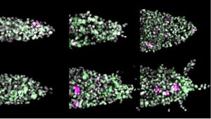
Fluorescence images of sample tumors show invasions into surrounding tissue over several days. Branching invasions are most pronounced in the lower right frame. (Photo illustration from image provided by the researchers/Princeton University.)
Credit:Princeton University

Other diets showed limited impact in the model

The study also tested tumor responses under different nutrient conditions, including high insulin, glycerol, and ketones.

These conditions showed little difference from baseline tumors, which remained relatively compact and did not develop invasive structures.

One unexpected result involved a simulated ketogenic diet, which is typically high in fat and low in carbohydrates.

Researchers had expected it might slow tumor progression. Instead, the tumors did not show improved outcomes compared to baseline conditions within the model.

“We were expecting a ketogenic diet to be protective,” Nelson said. “Yet we didn’t see that here.”

She added that the model may not fully capture the complexity of how such diets interact with the human body, particularly immune responses and other systemic factors.

Study highlights limits of lab-based cancer models

The findings are based on 3D microfluidic tumor models, which aim to replicate aspects of real biological environments more accurately than traditional lab methods.

Unlike 2D cell cultures, which grow in simplified conditions, these models simulate both the physical and chemical environment of tumors. At the same time, they remain more controlled than animal studies, allowing researchers to isolate specific variables such as diet.

Even so, the researchers caution that the results are limited.

The tumors studied are simplified representations and do not include the full range of interactions present in the human body. That means the findings cannot be directly translated into dietary recommendations or clinical outcomes.

“Every tumor is an individual’s tumor,” Nelson said, noting the challenge of capturing the full diversity of cancer behavior in a single study.

New direction for diet and cancer research

The study adds to a growing body of evidence suggesting that diet influences cancer progression, though the mechanisms remain unclear.

By focusing on structural changes rather than growth rates, the Princeton team offers a different lens for understanding how nutrients interact with tumors.

The research also points to potential targets for future therapies, particularly genes involved in tissue breakdown and tumor invasion.

For now, the findings remain an early step.

They show how fat may change the way tumors behave under controlled conditions, opening new questions about how those processes unfold inside the human body.

Also Read:

New MetaRing Sensor Spots Breast Cancer Drug Response in 10 Minutes

Researchers at the Chinese Academy of Sciences have developed a biosensor that can quickly detect how breast cancer responds to paclitaxel. The system, called MetaRing, analyzes tiny biological samples and delivers results in about 10 minutes. The study, published recently, points to faster, more personalized chemotherapy decisions.


Not just chemotherapy, now phototherapy is here for cancer treatment [Details]

One approach to treating cancer is photodynamic therapy using photo-uncaging systems, in which light is used to activate a cancer-fighting agent in situ at the tumor. However, suitable agents must be stable under visible light, have an anti-tumor effect in low-oxygen environments, and have the ability to be activated by low-energy tissue-penetrative red light – a combination of properties that is difficult to achieve. Now, a team from The Institute of Industrial Science at The University of Tokyo has developed a new platform that uses, for the first time, organorhodium(III) phthalocyanine complexes to achieve this combination of traits.

Conventional photodynamic techniques depend on the formation of reactive oxygen species to destroy tumor cells, but many tumors contain environments that lack oxygen. Photo-uncaging systems, where the agent is administered in an inactive form and then activated, or “uncaged”, in the location of the tumor, address this issue. They uncage alkyl radicals, which are known to be capable of inducing cell death both with and without the presence of oxygen. Alkyl radicals are converted into terminal aldehydes in the presence of oxygen, and these terminal aldehydes can also induce cell death. The team used molecules called “organorhodium(III) phthalocyanine (Pc) complexes” to develop, for the first time, a novel platform for photo-uncaging therapy.

Researchers from The Institute of Industrial Science, The University of Tokyo have developed a streamlined photo-uncaging system for photodynamic cancer therapy, using a pulse of light for tumor-specific activation of a cancer-fighting agent/CREDIT
Institute of Industrial Science, The University of Tokyo

“The organorhodium(III) phthalocyanine (Pc) complexes we developed are highly stable under ambient light during the processes of synthesis, purification, and measurement, but can be activated by a laser that gives out nanosecond pulses of red light,” explains lead author Kei Murata. These nanosecond-pulsing lasers (pulsing for a billionth of a second) are relatively easy for medical staff to handle.

They went on to show that the compounds that were released after the organorhodium(III) phthalocyanine (Pc) complexes were activated showed toxicity to HeLa cells, a cell line developed from cancer, indicating that these compounds would have the ability to fight cancer if released inside a tumor.

“Our new technology could allow the photochemical generation of a wide variety of alkyl radicals and aldehydes, making possible the site-selective release of various bioactive molecules,” says senior author Kazuyuki Ishii. As an improvement on other photo-uncaging systems, it opens an exciting new avenue for the treatment of cancer by phototherapy.

Ovarian cancer detection takes a step forward with liquid biopsy

Ovarian cancer is most often found in people of middle-age or older as the Wilmot study showed that the mean age of participants was 56.

Of the 183 participants, 42 were found to have ovarian cancer, which is 23 percent. The technology also discovered that 20 other participants had non-ovarian cancers.

Ovarian cancer symptoms can be vague, such as gas and bloating, but there are some that should not be ignored, Moore said: pelvic pain or pressure, feeling full quickly after eating, vaginal discharge or abnormal bleeding, urgency to urinate frequently, fatigue, upset stomach, pain during sex, constipation, or menstrual changes.

Because ovarian cancer is most often diagnosed in later stages.

A new type of technology can capture stray ovarian cancer cells from a simple blood test and successfully predict cancer in people who have a lesion or cyst in the pelvic region, according to a new study by a Wilmot Cancer Institute physician/scientist.

Nearly 200 local people participated in the study.

One of those local participants, Toni Masci, 51, of Fairport, took part in Moore’s study by providing blood samples for analysis. She had an ovarian cyst that burst — only to find out that a large tumor was also in her abdomen. She was treated with surgery and six rounds of chemotherapy in 2017 for stage 1 ovarian cancer, and just celebrated the milestone of five years in remission.

“I feel lucky to be part of this,” Masci said. “As most people know, ovarian cancer usually doesn’t get detected early. If Dr. Moore hadn’t been doing this research, we might not have had this advance and I might not be here.”

Currently, there is no routine ovarian cancer screening method available for people who do not have symptoms or a known lesion. And yet, the new technology, called a “liquid biopsy,” developed by United Kingdom-based ANGLE PLC, and the URMC team at Wilmot, advances the field in a couple of important ways, according to the study:

  • It confirmed for physicians quickly and accurately that cancer was present in patients who were scheduled for surgery or other procedures. The detection enabled physicians to classify which patients needed immediate care from a specially trained gynecological oncologist to improve survival.
  • The study analyzed gene expression from captured cells in blood and evaluated 72 different gene transcripts and seven blood biomarkers related to ovarian cancer (including CA125). From this collection, the study identified nine gene transcripts and four biomarkers that were useful for detecting cancers. They were used to develop an algorithm known as MAGIC (Malignancy Assessment using Gene Identification in Captured Cells). The algorithm achieved a sensitivity of 95 percent and an accuracy of 83 percent for detecting ovarian cancer.
  • In the clinical trial, MAGIC also was able to detect ovarian cancer in early and late stages. Early-stage detection is critical for survival and difficult to achieve. And, the test picked up other types of cancer that had spread to the pelvic region or originated there.

“This is an important step forward for the detection of ovarian cancer in patients with a pelvic mass,” Moore said. “The fact that we can capture circulating tumor cells and analyze them from a simple blood draw is extremely exciting.”

Being able to find circulating tumor cells is the key, Moore said. These are rare, living cells that break off from the original tumor. They have an estimated ratio in the blood of one in 100 million to one in one billion. The technology captures the rare cells and allows for genetic analysis in a single tool within a couple of hours.


Currently, if a person has a suspicious lesion, surgery is necessary to diagnose ovarian cancer, and annually, more than 200,000 people in the U.S. are in this situation. A non-invasive test that predicts malignancy beforehand would enable people with the highest risk to have surgery done by an oncology specialist with greater experience and surgical volume for these types of cases, Moore said.

Masci, a U.S. Navy veteran and esthetician at a local salon, was 46 years old in January 2017 when her cancer was diagnosed.

“I was in such shock,” she said. “Looking back, I did have some symptoms: bloating, my back hurt, weight loss, and when I would sit down to eat I would feel full right away.”

She enrolled in the study a month later, and Moore performed her ovarian cancer surgery.

“I had wonderful care from everyone at Wilmot,” Masci added, “but I can’t say enough good things about Dr. Moore. He needs to clone himself a million times.”

ANGLE Europe Limited funded the study. Moore has worked extensively with the company to test its detection system. Earlier this summer, the FDA gave approval for the same tool to be used to track breast cancer cells that have spread. Moore’s lab was the sole location nationally to test the reproducibility of the breast cancer tests, and local residents were also involved in that clinical trial.


Healthcard for Cancer Treatment

Rashtriya Swasthya Bima Yojana (RSBY), a centrally sponsored scheme, provides health insurance coverage to Below Poverty Line (BPL) families and including other 11 categories of Unorganized Workers (UOWs) who are enrolled under the scheme.

Senior Citizen Health Insurance Scheme (SCHIS) is also implemented w.e.f. 01.04.2016. Under this, health coverage is available for Rs.30,000/- per annum per senior citizen for treatment packages, over and above RSBY entitlement.

Each family enrolled in the scheme is entitled for hospitalization benefits in Government empanelled hospitals (including both private and public). RSBY and SCHIS cover oncology treatment within prescribed benefits ceiling.

Under comprehensive Primary Healthcare, operational guidelines for Prevention, Screening and Control of Common Non-Communicable Diseases: Hypertension, Diabetes and Common Cancers (Oral, Breast, Cervix) have been issued under National Health Mission which envisage preparation of Health Cards for individuals above 30 years of age. These Cards primarily will be health records of individuals listing health issues/diseases/disabilities and exposures to risk factors of common NCDs including cancer.

The Minister of State (Health and Family Welfare), Smt Anupriya Patel stated this in a written reply in the Rajya Sabha here today.