Not just chemotherapy, now phototherapy is here for cancer treatment [Details]

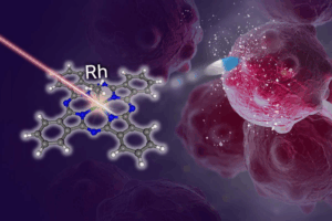
One approach to treating cancer is photodynamic therapy using photo-uncaging systems, in which light is used to activate a cancer-fighting agent in situ at the tumor. However, suitable agents must be stable under visible light, have an anti-tumor effect in low-oxygen environments, and have the ability to be activated by low-energy tissue-penetrative red light – a combination of properties that is difficult to achieve. Now, a team from The Institute of Industrial Science at The University of Tokyo has developed a new platform that uses, for the first time, organorhodium(III) phthalocyanine complexes to achieve this combination of traits.

Conventional photodynamic techniques depend on the formation of reactive oxygen species to destroy tumor cells, but many tumors contain environments that lack oxygen. Photo-uncaging systems, where the agent is administered in an inactive form and then activated, or “uncaged”, in the location of the tumor, address this issue. They uncage alkyl radicals, which are known to be capable of inducing cell death both with and without the presence of oxygen. Alkyl radicals are converted into terminal aldehydes in the presence of oxygen, and these terminal aldehydes can also induce cell death. The team used molecules called “organorhodium(III) phthalocyanine (Pc) complexes” to develop, for the first time, a novel platform for photo-uncaging therapy.

Phototherapy

Researchers from The Institute of Industrial Science, The University of Tokyo have developed a streamlined photo-uncaging system for photodynamic cancer therapy, using a pulse of light for tumor-specific activation of a cancer-fighting agent/CREDIT
Institute of Industrial Science, The University of Tokyo

“The organorhodium(III) phthalocyanine (Pc) complexes we developed are highly stable under ambient light during the processes of synthesis, purification, and measurement, but can be activated by a laser that gives out nanosecond pulses of red light,” explains lead author Kei Murata. These nanosecond-pulsing lasers (pulsing for a billionth of a second) are relatively easy for medical staff to handle.

They went on to show that the compounds that were released after the organorhodium(III) phthalocyanine (Pc) complexes were activated showed toxicity to HeLa cells, a cell line developed from cancer, indicating that these compounds would have the ability to fight cancer if released inside a tumor.

“Our new technology could allow the photochemical generation of a wide variety of alkyl radicals and aldehydes, making possible the site-selective release of various bioactive molecules,” says senior author Kazuyuki Ishii. As an improvement on other photo-uncaging systems, it opens an exciting new avenue for the treatment of cancer by phototherapy.

Ovarian cancer detection takes a step forward with liquid biopsy

Ovarian cancer is most often found in people of middle-age or older as the Wilmot study showed that the mean age of participants was 56.

Of the 183 participants, 42 were found to have ovarian cancer, which is 23 percent. The technology also discovered that 20 other participants had non-ovarian cancers.

Ovarian cancer symptoms can be vague, such as gas and bloating, but there are some that should not be ignored, Moore said: pelvic pain or pressure, feeling full quickly after eating, vaginal discharge or abnormal bleeding, urgency to urinate frequently, fatigue, upset stomach, pain during sex, constipation, or menstrual changes.

Because ovarian cancer is most often diagnosed in later stages.

A new type of technology can capture stray ovarian cancer cells from a simple blood test and successfully predict cancer in people who have a lesion or cyst in the pelvic region, according to a new study by a Wilmot Cancer Institute physician/scientist.

Nearly 200 local people participated in the study.

One of those local participants, Toni Masci, 51, of Fairport, took part in Moore’s study by providing blood samples for analysis. She had an ovarian cyst that burst — only to find out that a large tumor was also in her abdomen. She was treated with surgery and six rounds of chemotherapy in 2017 for stage 1 ovarian cancer, and just celebrated the milestone of five years in remission.

“I feel lucky to be part of this,” Masci said. “As most people know, ovarian cancer usually doesn’t get detected early. If Dr. Moore hadn’t been doing this research, we might not have had this advance and I might not be here.”

Currently, there is no routine ovarian cancer screening method available for people who do not have symptoms or a known lesion. And yet, the new technology, called a “liquid biopsy,” developed by United Kingdom-based ANGLE PLC, and the URMC team at Wilmot, advances the field in a couple of important ways, according to the study:

  • It confirmed for physicians quickly and accurately that cancer was present in patients who were scheduled for surgery or other procedures. The detection enabled physicians to classify which patients needed immediate care from a specially trained gynecological oncologist to improve survival.
  • The study analyzed gene expression from captured cells in blood and evaluated 72 different gene transcripts and seven blood biomarkers related to ovarian cancer (including CA125). From this collection, the study identified nine gene transcripts and four biomarkers that were useful for detecting cancers. They were used to develop an algorithm known as MAGIC (Malignancy Assessment using Gene Identification in Captured Cells). The algorithm achieved a sensitivity of 95 percent and an accuracy of 83 percent for detecting ovarian cancer.
  • In the clinical trial, MAGIC also was able to detect ovarian cancer in early and late stages. Early-stage detection is critical for survival and difficult to achieve. And, the test picked up other types of cancer that had spread to the pelvic region or originated there.

“This is an important step forward for the detection of ovarian cancer in patients with a pelvic mass,” Moore said. “The fact that we can capture circulating tumor cells and analyze them from a simple blood draw is extremely exciting.”

Being able to find circulating tumor cells is the key, Moore said. These are rare, living cells that break off from the original tumor. They have an estimated ratio in the blood of one in 100 million to one in one billion. The technology captures the rare cells and allows for genetic analysis in a single tool within a couple of hours.


Currently, if a person has a suspicious lesion, surgery is necessary to diagnose ovarian cancer, and annually, more than 200,000 people in the U.S. are in this situation. A non-invasive test that predicts malignancy beforehand would enable people with the highest risk to have surgery done by an oncology specialist with greater experience and surgical volume for these types of cases, Moore said.

Masci, a U.S. Navy veteran and esthetician at a local salon, was 46 years old in January 2017 when her cancer was diagnosed.

“I was in such shock,” she said. “Looking back, I did have some symptoms: bloating, my back hurt, weight loss, and when I would sit down to eat I would feel full right away.”

She enrolled in the study a month later, and Moore performed her ovarian cancer surgery.

“I had wonderful care from everyone at Wilmot,” Masci added, “but I can’t say enough good things about Dr. Moore. He needs to clone himself a million times.”

ANGLE Europe Limited funded the study. Moore has worked extensively with the company to test its detection system. Earlier this summer, the FDA gave approval for the same tool to be used to track breast cancer cells that have spread. Moore’s lab was the sole location nationally to test the reproducibility of the breast cancer tests, and local residents were also involved in that clinical trial.


Boosting physical activity/curbing sitting time likely to lower breast cancer risk:Mendelian randomisation study reveals

Boosting physical activity levels and curbing sitting time are highly likely to lower breast cancer risk, finds research designed to strengthen proof of causation and published online in the British Journal of Sports Medicine.

The findings were generally consistent across all types and stages of the disease, reveals the Mendelian randomisation study, prompting the researchers to recommend a stronger focus on exercise as a way of warding off breast cancer.

Mendelian randomisation is a technique that uses genetic variants as proxies for a particular risk factor—in this case lifelong physical activity levels/sedentary behaviour—to obtain genetic evidence in support of a causal relationship.

Exercise/Photo:en.wikipedia.org

Observational studies show that physical inactivity and sedentary behaviour are linked to higher breast cancer risk, but proving they cause breast cancer is another matter.

The researchers therefore used Mendelian randomisation to assess whether lifelong physical activity and sitting time might be causally related to breast cancer risk in general, and specifically to different types of tumour.

They included data from 130,957 women of European ancestry: 69, 838 of them had tumours that had spread locally (invasive); 6667 had tumours that hadn’t yet done so (in situ); and a comparison group of 54,452 women who didn’t have breast cancer.

Exercise-Yoga/Photo:en.wikipedia.org

The researchers then drew on previously published studies that had used the vast repository of UK Biobank data on potential genetic explanations for overall predisposition to physical activity, vigorous physical activity, or sitting time—as measured by wrist-worn activity trackers—to genetically predict how physically active or inactive their own study participants were.

Next, the researchers estimated overall breast cancer risk, according to whether the women had or hadn’t gone through the menopause; and by cancer type,stage (size and extent of tumour spread), and grade (degree of tumour cell abnormality).

Analysis of the data showed that a higher overall level of genetically predicted physical activity was associated with a 41% lower risk of invasive breast cancer, and this was largely irrespective of menopausal status, tumour type, stage, or grade.

Similarly, genetically predicted vigorous physical activity on 3 or more days of the week was associated with a 38% lower risk of breast cancer, compared with no self-reported vigorous activity. These findings were consistent across most of the case groups.

cancer cells/photo:en.wikipedia.org

Finally, a greater level of genetically predicted sitting time was associated with a 104% higher risk of triple negative breast cancer. These findings were consistent across hormone-negative tumour types.

The findings were unchanged after factoring in the production by a single gene of two or more apparently unrelated effects (pleiotropy), such as smoking and overweight, for example.

There are plausible biological explanations for their findings, say the researchers, who point to a reasonable body of evidence indicating numerous causal pathways between physical activity and breast cancer risk, such as overweight/obesity, disordered metabolism, sex hormones, and inflammation.

“Mechanisms linking sedentary time and cancer are likely to at least partially overlap with those underpinning the physical activity relationship,” suggest the researchers.

Their findings provide “strong evidence” that more overall physical activity and less sitting time are likely to reduce breast cancer risk, they say.

And they conclude: “Increasing physical activity and reducing sedentary time are already recommended for cancer prevention. Our study adds further evidence that such behavioural changes are likely to lower the incidence of future breast cancer rates.

“A stronger cancer-control focus on physical activity and sedentary time as modifiable cancer risk factors is warranted, given the heavy burden of disease attributed to the most common cancer in women.”