‘Mystery gene’ matures the skeleton of the cell

“I’m a professional pin-in-a-haystack seeker,” geneticist Thijn Brummelkamp responds when asked why he excels at tracking down proteins and genes that other people did not find, despite the fact that some have managed to remain elusive for as long as forty years. His research group at the Netherlands Cancer Institute has once again managed to track down one of these “mystery genes” – the gene that ensures that the final form of the protein actin is created, a main component of our cell skeleton.

Cell biologists are very interested in actin, because actin – a protein of which we produce more than 100 kilograms in our lifetime – is a main component of the cell skeleton and one of the most abundant molecules in a cell. Large quantities can be found in every cell type and it has many purposes: it gives shape to the cell and makes it firmer, it plays an important role in cell division, it can propel cells forward, and provides strength to our muscles. People with faulty actin proteins often suffer from muscle disease. Much is known about the function of actin, but how the final form of this important protein is made and which gene is behind it? “We didn’t know,” says Brummelkamp, whose mission is to find out the function of our genes.

Multi-purpose method for genetics in human cells
Together with other researchers, Brummelkamp uses this multi-purpose method to find the genetic causes of particular conditions. He has already shown how the Ebola virus and a number of other viruses, as well as certain forms of chemotherapy, manage to enter a cell. He also investigated why cancer cells are resistant to certain types of therapy and discovered a protein found in cancer cells that acts as a brake on the immune system. This time he went looking for a gene that matures actin – and as a result, the skeleton of the cell.

Microscopy image of actine. (Actine is yellow, cell core is blue)

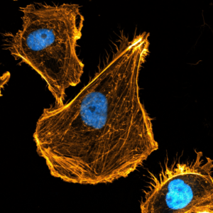
Microscopy image of actine. (Actine is yellow, cell core is blue)/CREDIT:Peter Haarh, Netherlands Cancer Institute

In search of scissors
Before a protein is completely “finished” – or mature, as the researchers describe it in Science – and can fully perform its function in the cell, it usually has to be stripped of a specific amino acid first. This amino acid is then cut from a protein by a pair of molecular scissors. This is also what occurs with  actin. It was known on which side of the actin the relevant amino acid is cut off. However, no one managed to find the enzyme that acts as scissors in this process.

Peter Haahr, postdoc in Brummelkamp’s group, worked on the following experiment: first he caused random mutations (mistakes) in random haploid cells. Then he selected the cells containing the immature actin by adding a fluorescently labeled antibody to his cells that fit in the exact spot where the amino acid is cut off. As a third and final step, he investigated which gene mutated after this process.

They called it ‘ACTMAP’
Then came the “eureka”-moment: Haahr had traced down the molecular scissors that cut the essential amino acid from actin. Those scissors turned out to be controlled by a gene with a previously unknown function; one no researcher had ever worked with. This means that the researchers were able to name the gene themselves, and they settled on ACTMAP (ACTin MAturation Protease).

More scissors found in the skeleton of the cell
ACTMAP is not the first mystery gene discovered by Brummelkamp that plays a role in our cell skeleton function. Using the same method, his group has been able to detect three unknown molecular scissors over recent years that cut an amino acid from tubulin, the other main component of the cell skeleton. These scissors allow tubulin to perform its dynamic functions properly inside the cell. The last scissors (MATCAP) were discovered and described in Science this year. Through this earlier work on the cell skeleton, Brummelkamp managed to arrive at actin.

Also Read:

Not just chemotherapy, now phototherapy is here for cancer treatment [Details]

Ovarian cancer detection takes a step forward with liquid biopsy

Hunger-controlling brain cells may offer path for new obesity drugs

Boosting physical activity/curbing sitting time likely to lower breast cancer risk:Mendelian randomisation study reveals

Boosting physical activity levels and curbing sitting time are highly likely to lower breast cancer risk, finds research designed to strengthen proof of causation and published online in the British Journal of Sports Medicine.

The findings were generally consistent across all types and stages of the disease, reveals the Mendelian randomisation study, prompting the researchers to recommend a stronger focus on exercise as a way of warding off breast cancer.

Mendelian randomisation is a technique that uses genetic variants as proxies for a particular risk factor—in this case lifelong physical activity levels/sedentary behaviour—to obtain genetic evidence in support of a causal relationship.

Exercise/Photo:en.wikipedia.org

Observational studies show that physical inactivity and sedentary behaviour are linked to higher breast cancer risk, but proving they cause breast cancer is another matter.

The researchers therefore used Mendelian randomisation to assess whether lifelong physical activity and sitting time might be causally related to breast cancer risk in general, and specifically to different types of tumour.

They included data from 130,957 women of European ancestry: 69, 838 of them had tumours that had spread locally (invasive); 6667 had tumours that hadn’t yet done so (in situ); and a comparison group of 54,452 women who didn’t have breast cancer.

Exercise-Yoga/Photo:en.wikipedia.org

The researchers then drew on previously published studies that had used the vast repository of UK Biobank data on potential genetic explanations for overall predisposition to physical activity, vigorous physical activity, or sitting time—as measured by wrist-worn activity trackers—to genetically predict how physically active or inactive their own study participants were.

Next, the researchers estimated overall breast cancer risk, according to whether the women had or hadn’t gone through the menopause; and by cancer type,stage (size and extent of tumour spread), and grade (degree of tumour cell abnormality).

Analysis of the data showed that a higher overall level of genetically predicted physical activity was associated with a 41% lower risk of invasive breast cancer, and this was largely irrespective of menopausal status, tumour type, stage, or grade.

Similarly, genetically predicted vigorous physical activity on 3 or more days of the week was associated with a 38% lower risk of breast cancer, compared with no self-reported vigorous activity. These findings were consistent across most of the case groups.

cancer cells/photo:en.wikipedia.org

Finally, a greater level of genetically predicted sitting time was associated with a 104% higher risk of triple negative breast cancer. These findings were consistent across hormone-negative tumour types.

The findings were unchanged after factoring in the production by a single gene of two or more apparently unrelated effects (pleiotropy), such as smoking and overweight, for example.

There are plausible biological explanations for their findings, say the researchers, who point to a reasonable body of evidence indicating numerous causal pathways between physical activity and breast cancer risk, such as overweight/obesity, disordered metabolism, sex hormones, and inflammation.

“Mechanisms linking sedentary time and cancer are likely to at least partially overlap with those underpinning the physical activity relationship,” suggest the researchers.

Their findings provide “strong evidence” that more overall physical activity and less sitting time are likely to reduce breast cancer risk, they say.

And they conclude: “Increasing physical activity and reducing sedentary time are already recommended for cancer prevention. Our study adds further evidence that such behavioural changes are likely to lower the incidence of future breast cancer rates.

“A stronger cancer-control focus on physical activity and sedentary time as modifiable cancer risk factors is warranted, given the heavy burden of disease attributed to the most common cancer in women.”

Air pollution is more dangerous for women than men: Study

The impact of breathing diesel exhaust fumes may be more severe for females than males, according to new research that will be presented at the European Respiratory Society International Congress in Barcelona, Spain [1].

Researchers looked for changes in people’s blood brought about by exposure to diesel exhaust. In both females and males, they found changes in components of the blood related to inflammation, infection and cardiovascular disease, but they found more changes in females than males.

The research was presented by Dr Hemshekhar Mahadevappa, from the University of Manitoba, Winnipeg, Canada and was a collaboration between two research groups led by Professor Neeloffer Mookherjee at the University of Manitoba and Professor Chris Carlsten at the University of British Columbia, Vancouver, Canada. Dr Mahadevappa told the Congress: “We already know that there are sex differences in lung diseases such as asthma and respiratory infections.

Air pollution/Photo:en.wikipedia.org

Research showed that breathing diesel exhaust creates inflammation in the lungs and has an impact on how the body deals with respiratory infections. In this study, we wanted to look for any effects in the blood and how these differ in females and males.”

The study involved ten volunteers, five female and five male, who were all healthy non-smokers. Each volunteer spent four hours breathing filtered air and four hours breathing air containing diesel exhaust fumes at three different concentrations – 20, 50 and 150 micrograms of fine particulate matter (PM2.5) per cubic metre – with a four-week break in between each exposure.

Volunteers donated blood samples 24 hours after each exposure and the researchers made detailed examinations of the volunteers’ blood plasma. Plasma is the liquid component of the blood that carries blood cells as well as hundreds of proteins and other molecules around the body. Using a well-established analysis technology called liquid chromatography–mass spectrometry, the researchers looked for changes in the levels of different proteins following exposure to diesel exhaust and compared the changes in females and males.

Airpollution/Photo:en.wikipedia.org

Comparing the plasma samples, the researchers found levels of 90 proteins that were distinctly different between female and male volunteers following exposure to diesel exhaust. Among the proteins that differed between females and males, were some that are known to play a role in inflammation, damage repair, blood clotting, cardiovascular disease and the immune system. Some of these differences became clearer when volunteers were exposed to the higher levels of diesel exhaust.

Professor Mookherjee explained: “These are preliminary findings, however they show that exposure to diesel exhaust has different effects in female bodies compared to male and that could indicate that air pollution is more dangerous for females than males.

“This is important as respiratory diseases such as asthma are known to effect females and males differently, with females more likely to suffer severe asthma that does not respond to treatments. Therefore, we need to know a lot more about how females and males respond to air pollution and what this means for preventing, diagnosing and treating their respiratory disease.”

Air pollution/photo:en.wikipedia.org

Professor Zorana Andersen from the University of Copenhagen, Denmark, is Chair of the European Respiratory Society Environment and Health Committee and was not involved in the research. She said: “We know that exposure to air pollution, especially diesel exhaust, is a major risk factor in diseases such as asthma and chronic obstructive pulmonary disease. There is very little we can do as individuals to avoid beathing polluted air, so we need governments to set and enforce limits on air pollutants.


Being away from excessive blue light from our gadgets slows ageing process

Changes in cell chemistry from blue light exposure observed in fruit flies could also potentially cause detrimental effects on our body, including accelerated aging.

Too much screen use has been linked to obesity and psychological problems. Now a new study has identified a new problem – a study in fruit flies suggests our basic cellular functions could be impacted by the blue light emitted by these devices. These results are published in Frontiers in Aging.

“Excessive exposure to blue light from everyday devices, such as TVs, laptops, and phones, may have detrimental effects on a wide range of cells in our body, from skin and fat cells, to sensory neurons,” said Dr Jadwiga Giebultowicz, a professor at the Department of Integrative Biology at Oregon State University and senior author of this study. “We are the first to show that the levels of specific metabolites –  chemicals that are essential for cells to function correctly – are altered in fruit flies exposed to blue light.“

“Our study suggests that avoidance of excessive blue light exposure may be a good anti-aging strategy,” advised Giebultowicz.

Mobile Phone/Photo:indiainternationaltimes

Turn off the light

The researchers at Oregon State University have previously shown that fruit flies exposed to light ‘turn on’ stress protective genes, and that those kept in constant darkness lived longer.

“To understand why high-energy blue light is responsible for accelerating aging in fruit flies, we compared the levels of metabolites in flies exposed to blue light for two weeks to those kept in complete darkness,” explained Giebultowicz.

Blue light exposure caused significant differences in the levels of metabolites measured by the researchers in the cells of fly heads. In particular, they found that the levels of the metabolite succinate were increased, but glutamate levels were lowered.

“Succinate is essential for producing the fuel for the function and growth of each cell. High levels of succinate after exposure to blue light can be compared to gas being in the pump but not getting into the car,” said Giebultowicz. “Another troubling discovery was that molecules responsible for communication between neurons, such as glutamate, are at the lower level after blue light exposure.”

Accelerating aging

The changes recorded by the researchers suggest that the cells are operating at suboptimal level, and this may cause their premature death, and further, explain their previous findings that blue light accelerates aging.

“LEDs have become the main illumination in display screens such as phones, desktops and TVs, as well as ambient lighting, so humans in advanced societies are exposed to blue light through LED lighting during most of their waking hours. The signaling chemicals in the cells of flies and humans are the same, so the there is potential for negative effects of blue light on humans,“ explains Giebultowicz.

Future work hopes to study the effects directly on human cells.

“We used a fairly strong blue light on the flies – humans are exposed to less intense light, so cellular damage may be less dramatic. The results from this study suggests that future research involving human cells is needed to establish the extent to which human cells may show similar changes in metabolites involved in energy production in response to excessive exposure to blue light,“ concluded Giebultowicz.