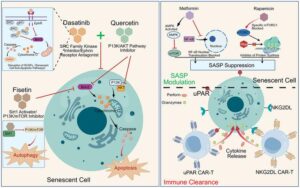
Mechanistic landscape of pro-longevity strategies. This figure illustrates the mechanistic action of key small-molecule drugs in clearing senescent cells, alongside immunotherapy strategies designed for the same purpose. Furthermore, it depicts the mechanisms by which anti-aging agents, such as SASP inhibitors, reduce the secretion of the senescence-associated secretory phenotype to achieve therapeutic anti-aging effects.
Credit Creative Commons Attribution License (CC BY 4.0),
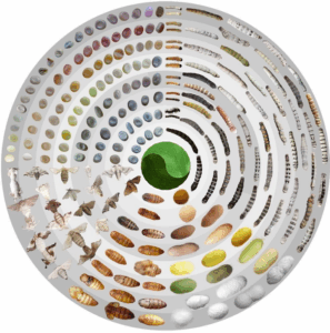
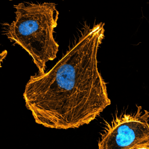
The paper stresses that senescent cells are not a single uniform group. Depending on their biological environment, some may help prevent fibrosis and support regeneration, while others promote inflammation, tissue degeneration, metabolic disorders and cancer development.
This growing understanding has led scientists to rethink anti-aging therapies. Instead of broadly eliminating all senescent cells, researchers are increasingly exploring “precision geroprotection” — a strategy aimed at selectively targeting harmful senescent cells while preserving beneficial ones important for tissue stability and recovery.
“Based on these insights, this review summarizes the induction mechanisms of cellular senescence and the subsequent evolution of their functional phenotypes across diverse tissues,” the authors wrote.
The review also outlines emerging anti-aging treatments. Early senolytic drugs such as Dasatinib, Quercetin and Fisetin were developed to destroy senescent cells by disrupting their survival pathways.
Newer approaches now include advanced immunotherapies such as CAR-T cell therapies targeting senescence-associated markers, along with “senomorphic” treatments that suppress harmful inflammatory signalling without killing the cells themselves.
The authors said technologies such as single-cell omics, lineage tracing and spatial profiling could help scientists better identify different senescent cell subtypes and develop safer, more precise therapies.
At the same time, the review warns that major clinical challenges remain. These include the lack of reliable biomarkers for senescent cells, difficulties in delivering targeted drugs, risks of unintended tissue damage and limited understanding of how senescent cells evolve over time in different organs.
The researchers cautioned that removing senescent cells indiscriminately could interfere with tissue repair, immune defence and structural stability in organs such as the brain, heart and lungs.
Overall, the review presents a more balanced understanding of cellular aging, arguing that future anti-aging medicine will likely depend on carefully tailored interventions rather than broad elimination of senescent cells.
Also Read:
Is your brain aging faster than you are? What holds the key
As a lifetime passes in front of our eyes, here’s the structure of how aging plays out