Evidence that babies react to taste, smell in the womb; Carrot for “laughter-face” response, kale for “cry-face” response: Study

A study led by Durham University’s Fetal and Neonatal Research Lab, UK, took 4D ultrasound scans of 100 pregnant women to see how their unborn babies responded after being exposed to flavours from foods eaten by their mothers.

Researchers looked at how the fetuses reacted to either carrot or kale flavours just a short time after the flavours had been ingested by the mothers.

Fetuses exposed to carrot showed more “laughter-face” responses while those exposed to kale showed more “cry-face” responses.

Their findings could further our understanding of the development of human taste and smell receptors.

The researchers also believe that what pregnant women eat might influence babies’ taste preferences after birth and potentially have implications for establishing healthy eating habits.

The study is published in the journal Psychological Science.

pregnant lady/Commons.wikimedia.org

Humans experience flavour through a combination of taste and smell. In fetuses it is thought that this might happen through inhaling and swallowing the amniotic fluid in the womb.

Lead researcher Beyza Ustun, a postgraduate researcher in the Fetal and Neonatal Research Lab, Department of Psychology, Durham University, said: “A number of studies have suggested that babies can taste and smell in the womb, but they are based on post-birth outcomes while our study is the first to see these reactions prior to birth.

“As a result, we think that this repeated exposure to flavours before birth could help to establish food preferences post-birth, which could be important when thinking about messaging around healthy eating and the potential for avoiding ‘food-fussiness’ when weaning.

“It was really amazing to see unborn babies’ reaction to kale or carrot flavours during the scans and share those moments with their parents.”

The research team, which also included scientists from Aston University, Birmingham, UK, and the National Centre for Scientific Research-University of Burgundy, France, scanned the mothers, aged 18 to 40, at both 32 weeks and 36 weeks of pregnancy to see fetal facial reactions to the kale and carrot flavours.

Mothers were given a single capsule containing approximately 400mg of carrot or 400mg kale powder around 20 minutes before each scan. They were asked not to consume any food or flavoured drinks one hour before their scans.

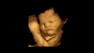
A 4D scan image of a fetus showing a neutral face/CREDIT: FETAP (Fetal Taste Preferences) Study, Fetal and Neonatal Research Lab, Durham University.

The mothers also did not eat or drink anything containing carrot or kale on the day of their scans to control for factors that could affect fetal reactions.

Facial reactions seen in both flavour groups, compared with fetuses in a control group who were not exposed to either flavour, showed that exposure to just a small amount of carrot or kale flavour was enough to stimulate a reaction.

Co-author Professor Nadja Reissland, head of the Fetal and Neonatal Research Lab, Department of Psychology, Durham University, supervised Beyza Ustun’s research. She said: “Previous research conducted in my lab has suggested that 4D ultrasound scans are a way of monitoring fetal reactions to understand how they respond to maternal health behaviours such as smoking, and their mental health including stress, depression, and anxiety.

“This latest study could have important implications for understanding the earliest evidence for fetal abilities to sense and discriminate different flavours and smells from the foods ingested by their mothers.”

Co-author Professor Benoist Schaal, of the National Centre for Scientific Research-University of Burgundy, France, said: “Looking at fetuses’ facial reactions we can assume that a range of chemical stimuli pass through maternal diet into the fetal environment.

This could have important implications for our understanding of the development of our taste and smell receptors, and related perception and memory.”

The researchers say their findings might also help with information given to mothers about the importance of taste and healthy diets during pregnancy.

They have now begun a follow-up study with the same babies post-birth to see if the influence of flavours they experienced in the womb affects their acceptance of different foods.

Research co-author Professor Jackie Blissett, of Aston University, said: “It could be argued that repeated prenatal flavour exposures may lead to preferences for those flavours experienced postnatally. In other words, exposing the fetus to less ‘liked’ flavours, such as kale, might mean they get used to those flavours in utero.

“The next step is to examine whether fetuses show less ‘negative’ responses to these flavours over time, resulting in greater acceptance of those flavours when babies first taste them outside of the womb.”

Related: http://dx.doi.org/10.1177/09567976221105460


New malaria analysis method reveals disease severity in minutes

Left untreated, malaria can progress from being mild to severe — and potentially fatal — in 24 hours. So researchers at the University of British Columbia developed a method to quickly and sensitively assess the progression of the mosquito-borne infectious disease, which remains a leading killer in low-income countries.

One way malaria wreaks havoc on the body is by causing excessive amounts of toxic heme, the non-protein component of hemoglobin, to accumulate in the bloodstream. Among other things, this free heme induces oxidative stress in red blood cells (RBCs), leading to their rigidification, destruction and subsequent removal from circulation — a condition known as hemolytic anemia.

In their study, which appeared in Integrative Biology, the UBC investigators found that RBCs become increasingly rigid in direct correlation with the concentration of oxidized heme, or hemin, in the blood. Since hemin is difficult to measure directly — it tends to insert itself into cell membranes — monitoring changes in RBC deformability can therefore serve as a reliable alternative marker of hemin-induced oxidative stress and malaria progression.

“Because this method is mechanical, it’s well suited for use in resource-poor countries, where the vast majority of malaria transmission takes place,” says Kerryn Matthews, a postdoctoral fellow at UBC and the study’s lead author. “Other methods of analyzing malaria severity require training or expensive equipment or chemicals that are not readily available in developing nations.”

To measure RBC deformability quickly and sensitively, the UBC investigators developed the “multiplex fluidic plunger”: a simple microfluidic device consisting of a parallel array of 34 funnel-shaped, micro-sized channels across which uniform, carefully controlled pressures can be simultaneously applied.

By loading the plunger with whole blood, docking an RBC at each channel and applying progressively higher pressures until the RBCs are squeezed through — or not, if an RBC is too stiff — one is able to determine the rigidity, or cortical tensions, of many cells at once and build an RBC deformability profile in minutes.

“The device can be easily integrated with a conventional microscope coupled with a digital camera,” says Matthews. “And the accompanying software, which does all the analysis and records the pressures, is simple to use.”

In addition to indicating the status of a malaria infection, RBC deformability information would be valuable in the development of antimalarial drugs, as well as in illuminating the mechanism by which RBCs are sequestered from circulation and destroyed.

Healthy RBCs are extremely flexible, capable of squeezing through spaces — the tiniest blood vessels, for example, or the channels between cells — that are just fractions of their original size. By rendering them less deformable, malaria parasites impair blood flow and ultimately cause organ failure and possibly death.

According to the World Health Organization, malaria killed an estimated 429,000 people and caused approximately 212 million clinical episodes in 2015. It primarily affects children and pregnant women in poor tropical and subtropical countries.

Scientists develop improved, potentially safer Zika vaccine

The worldwide Zika threat first emerged in 2015, infecting millions as it swept across the Americas. It struck great fear in pregnant women, as babies born with severe brain birth defects quickly overburdened hospitals and public health care systems.

In response, there has been a flurry of heroic scientific efforts to stop Zika. Whole governments, academic labs and pharmaceutical companies have raced to develop Zika vaccines ever since global health experts first realized the dangers wrought by the mosquito-borne virus.

Now, ASU has taken a major step forward in boosting Zika prevention efforts.

ASU Biodesign Institute scientist Qiang “Shawn” Chen has led his research team to develop the world’s first plant-based Zika vaccine that could be more potent, safer and cheaper to produce than any other efforts to date.

“Our vaccine offers improved safety and potentially lowers the production costs more than any other current alternative, and with equivalent effectiveness,” said Chen, a researcher in the Biodesign Center for IVV and professor in the School of Life Sciences. “We are very excited about these results.”

Rapid response network

Several potential Zika vaccines have had promising results in early animal and human tests. Last year, the Food and Drug Administration approved the first human testing of a Zika vaccine candidate, and this summer, a $100 million U.S. government-led clinical trial is underway.

But currently, there are no licensed vaccines or therapeutics available to combat Zika.

Several dedicated ASU scientists also heeded the call to action, wanting to use their special know-how to find a way to overcome the pandemic crisis.

First, ASU chemist Alexander Green, along with collaborators at Harvard, developed a more rapid and reliable Zika test, an achievement highlighted by Popular Science in its “Best of What’s New” of 2016.

Now, Chen may have come up with a better vaccine candidate based on a key Zika protein. Chen is a viral expert who has worked for the past decade on plant-based therapeutics and vaccines against West Nile virus and Dengue fever, which come from the same Zika family, called flaviviruses.

He honed in on developing a vaccine against a part of a Zika viral protein, called DIII, that plays a key role for the virus to infect people.

“All flaviviruses have the envelope protein on the outside part of the virus. It has three domains. “The domain III has a unique stretch of DNA for the Zika virus, and we exploited this to generate a robust and protective immune response that is unique for Zika,” said Chen.

They first grew the envelope protein in bacteria, then switched to prepare the DIII protein domain in tobacco plants.

After developing enough material for the new vaccine candidate, Chen’s team performed immunization experiments in mice, which induced antibody and cellular immune responses that have been shown to confer 100 percent protection against multiple Zika virus strains in a mouse challenge.

Producing plant-based vaccines, especially in tobacco plants, is old hat for ASU researchers like Chen. For more than a decade, they’ve been producing low-cost vaccines in plants to fight devastating infectious diseases in the developing world.

It’s the same approach ASU plant research pioneer Charles Arntzen used when he played a key role in developing ZMapp, the experimental treatment used during the Ebola outbreak.

Artntzen’s Biodesign colleagues, including Chen, Hugh Mason and Tsafrir Mor, have continued to pursue plant-based vaccines and therapeutics to combat West Nile virus, dengue fever, nerve agents and even cancer.

Effective but not foolproof

While Chen has been cheering on Zika vaccine progress from other researchers, in each case, there can be side effects.

To date, other scientists have tested several kinds of vaccines on mice –including one made from DNA and another from an inactivated form of the virus. With just one dose, both vaccines prompted the creation of antibodies that shielded the animals from becoming infected when they were exposed to the virus.

Any heat-killed vaccine runs the risk of accidentally injecting a live version of the virus if there is an error made in the vaccine production protocol. This tragic scenario happened occasionally with the polio vaccine.

For the second research group, they used the complete Zika envelope protein for their vaccine. Since envelope protein domains I and II are similar to West Nile and dengue viruses, this can cause a dangerous cross-reactive immune response.

“When you make the full native envelope protein as the basis for a vaccine, it will induce antibodies against DI, DII and the DIII domains of the protein,” explained Chen. “Those who have been prior exposed to DI and DII of other members of the Zika virus family may be prone to developing very bad symptoms, or in some cases, fatalities for dengue.”

In fact, animal experiments have shown that prior exposure to dengue or West Nile virus makes the Zika infection and symptoms much worse, suggesting a similar risk for people who had prior exposure to dengue (especially in South America, where it is more common).

“If you have prior exposure to dengue, and then have Zika exposure, the Zika infection may be much worse, and for men, may increase the likelihood of sexual transmission,” said Chen.

Chen’s protein-based vaccine uses the smallest and most unique part of the Zika virus that can still elicit a potent and robust immune response.

“In our approach, we make what we call a pseudovirus. It’s a fake virus. The pseudovirus displays only the DIII part of the envelope protein on the surface. This is at least as potent as previous vaccine versions.”

And he is very confident that his DIII-based protein vaccine will be safer.

“We did a test to make sure that the vaccine produces a potent protective immune response, but also, that it does not produce antibodies that may be cross reactive for dengue, West Nile, yellow fever or others,” said Chen.

Fast track to the clinic

During the height of the Zika pandemic, whole countries of women were told not to become pregnant, due to babies born with a severe brain defect called microcephaly, in which the head and brain don’t develop properly.

There have also been vision and hearing defects and learning disabilities associated with less severe infections.

To make matters worse, in adults, a debilitating nervous system condition called Gullian-Barre syndrome has also been shown to be caused by Zika.

While the most severe wave of the Zika pandemic has ebbed, it won’t go away anytime soon, and a vaccine still offers the best hope.

Tens of millions more could still be infected in the Americas in the coming years (see WHO fact sheet).

The ASU scientists were able to mobilize quickly from idea to proof-of-concept because they could leverage funds from an NIAID grant and seed funds from the Biodesign Institute.

These are all made possible by generous federal, state and public support, including sales tax generated from the long-time Arizona innovation booster, voter-approved Proposition 301.

“This is a great example of the brightest minds quickly coming together, with public support, to take on one of the most significant public health challenges of our time,” said Josh LaBaer, executive director of the Biodesign Institute.

“That’s the essence of Biodesign at its best, and we hope this important proof-of-principal of a Zika vaccine can be translated quickly into the clinic.”

With the successful proof-of-principle, Chen hopes to partner with the medical community to begin the first phase of a human clinical trial in the next two years.

“Above all, we have to ensure the utmost safety with any Zika vaccine, especially because the people who will need it most, pregnant women, have the most worries about their own health, and the health of the fetus,” said Chen. “This has to be 100 percent safe and effective.”

Along with Chen, the research team included Ming Yang, Huafang “Lily” Lai and Haiyan Sun.

The research was published in the online version of Scientific Reports.


Controlling Chronic Anaemia in Women

According to National Family Health Survey (NFHS)–IV (2015-16), the prevalence of anemia among women aged 15 to 49 years is 53%. In order to prevent anaemia among women and children, National Iron Plus Initiative (NIPI) has been launched in 2013, a flagship scheme under the National Health Mission based on the life-cycle approach, under which iron-folic acid supplementation is provided to the vulnerable age groups. It includes Pregnant and lactating women and Women in reproductive age (WRA) group.

Measures taken by Government to ensure optimum nutritional requirements for women are:

• Universal screening of pregnant women for anaemia is a part of ante-natal care and all pregnant women are to be provided iron and folic acid tablets during their ante-natal and post natal visits through the existing network of sub-centers and primary health centres and other health facilities as well as through outreach activities at Village Health & Nutrition Days (VHNDs) and also through the ASHAs. After the first trimester of pregnancy, every pregnant woman during ANC is to be given iron and folic acid (IFA) tablets for six months, and six months post-partum. Pregnant women, who are found to be clinically anaemic, are to be given double the dose of IFA.

• Government of India has given directions to the States for identification and tracking of severely anaemic cases at all the sub centres and PHCs for their timely management.

• Health and nutrition education to promote dietary diversification, inclusion of iron foliate rich food as well as food items that promote iron absorption.

• To address anaemia due to worm infestation, deworming of pregnant women is done after first trimester preferably in second trimester of pregnancy.

• To tackle the problem of anemia due to malaria particularly in pregnant women and children, Long Lasting Insecticide Nets (LLINs) and Insecticide Treated Bed Nets (ITBNs) are being distributed in endemic areas.

The Minister of State (Health and Family Welfare), Sh Faggan Singh Kulaste stated this in a written reply in the Rajya Sabha here today.