Chronic oral inflammation may impair female fertility by triggering a systemic immune response that affects the ovaries. A new study shows this leads to oxidative damage, reduced egg quality, disrupted follicle development and reduced live birth rate. These findings point to a potential biological link between oral health and unexplained infertility, opening new directions for future treatments.
[Hebrew University of Jerusalem]– A new study led by Prof. Michael Klutstein at the Hebrew University of Jerusalem and Prof. Asaf Wilensky at the Hebrew University-Hadassah Medical center and spearheaded by the students Dr. Paz Kles and Stephen Amehohas uncovered a striking biological link between chronic oral inflammation and female fertility, suggesting that conditions in the mouth may have far-reaching effects on reproductive health.
Published in the Journal of Dental Research, the study shows that persistent inflammation in the oral cavity can impair ovarian function, reduce egg quality, and ultimately lower fertility rates.
Researchers examined in a mouse model inflammation associated with dental implants, a common clinical scenario, and tracked how immune signals spread throughout the body. Their findings reveal that inflammation does not remain confined to the oral cavity but triggers a systemic immune response that reaches the ovaries.
The consequences were significant. Chronic oral inflammation in the animals was linked to increased levels of inflammatory cytokines in the ovaries, along with shifts in immune cell populations. This was accompanied by oxidative damage to ovarian tissue, impaired development of follicles, and reduced quality of oocytes.
These biological changes translated into measurable reproductive outcomes, with markedly reduced birth rates observed under inflammatory conditions in the animals.
The study also identified deeper cellular effects. Oocytes exhibited DNA damage and epigenetic alterations resembling those seen in reproductive aging, pointing to a possible mechanism by which inflammation accelerates the decline in fertility.
“Inflammation is often thought of as a localized response, but our findings show that it can have systemic consequences that extend as far as the reproductive system,” said Prof. Michael Klutstein. “This work suggests that chronic oral inflammation may be an underrecognized factor in female infertility, potentially contributing to cases that currently have no clear explanation.”
The findings add to growing evidence that oral health is closely linked to overall health. Chronic oral inflammatory conditions such as periodontitis are widespread and have already been associated with a range of systemic diseases.
The researchers note that further investigation in clinical settings will be essential to determine how these findings translate to patient care. If confirmed, the work could open new avenues for diagnosis and treatment, including the use of anti-inflammatory or antioxidant approaches to improve fertility outcomes.
A new study has found that children born to mothers lacking a specific fatty acid in their blood during pregnancy face a significantly higher risk of developing asthma in early life, shedding fresh light on how prenatal conditions shape long-term respiratory health.
Childhood asthma—often marked by shortness of breath, persistent coughing, and frequent respiratory infections—remains one of the most widespread chronic illnesses among children globally. Increasingly, scientists are tracing its origins back to the earliest stages of life, including the prenatal period.
Researchers from the University of Copenhagen and the Copenhagen Prospective Studies on Asthma in Childhood (COPSAC), based at the Danish Pediatric Asthma Center, examined the role of a fatty acid molecule known as 12-HETE in pregnant women. Their findings suggest that the absence of this molecule is associated with a markedly higher likelihood of children developing asthmatic bronchitis within their first year.
Over a ten-year observation period, the study found that children whose mothers lacked measurable levels of 12-HETE faced a 62 percent higher incidence of asthma compared to those whose mothers had detectable levels. However, researchers caution that the findings establish correlation rather than direct causation.
The study also identified broader health implications. Infants born to mothers without measurable 12-HETE were more prone to repeated respiratory infections and showed distinct differences in airway bacterial composition. These early-life changes in the microbiome and immune response are believed to contribute to the elevated asthma risk.
According to the researchers, differences in airway biology can be detected as early as one month after birth. Lower levels of the fatty acid during fetal development may impair immune system maturation, leading to an altered lung microbiome and increased vulnerability to infections.
The findings also add nuance to the role of omega-3 supplements during pregnancy. While earlier research has suggested that omega-3 intake—commonly through fish oil—can help reduce asthma risk in children, this study indicates that the benefit may depend on the presence of 12-HETE. Among mothers with measurable levels of the fatty acid, omega-3 supplementation was linked to a 58 percent reduction in early childhood asthma. No such effect was observed in mothers lacking the molecule.
This suggests that blanket supplementation strategies may not be equally effective for all pregnant women, and future interventions could be tailored based on individual biological markers.
Despite the promising insights, researchers stress that the findings are not yet ready for clinical application. More precise benchmarks for measuring 12-HETE levels are needed before it can be used to guide treatment or prevention strategies.
The research draws on data from two major cohorts—the Danish COPSAC2010 study and the US-based VDAART cohort—tracking more than 1,600 mothers and their children. The consistency of findings across both groups strengthens the reliability of the results, even as questions around causality remain unresolved.
Taken together, the study points to the potential of 12-HETE as a biomarker that could one day enable more personalised approaches to preventing childhood asthma—an area of growing urgency given the condition’s global prevalence.
In a significant international study, it has been observed that the health, life experience of fathers can have a significant effect on the pregnancy outcome and future well being of the children which argues the long held maternal-based approach to reproductive care.
The study, which was published in The Lancet and conducted by the team of scientists at the University of Southampton in collaboration with other international partners, claims that the prenatal health of men has long been disregarded despite its quantifiable effects on maternal health and child development.
Based on the evidence of biological, behavioural and social sciences, the study describes the ways in which the physical health of a father, his age, mental health, substance use and childhood experiences could influence the results of pregnancy. There are instances where scientists asserted that paternal factors can be as intense as -or even more so, than the conventional maternal.
Professor Keith Godfrey, a principal researcher of the study, indicated that the results represented a breakthrough in the comprehension of the role of parental health on the future generation. He mentioned that although the health of mothers is important, paying attention to it only leads to the neglect of a larger number of factors that trigger well before pregnancy.
The study presents a framework based on preconception health, in which the upbringing, education, environment of a man and exposure to stress can be influenced and impact on his health in reproductive years. The same factors, in their turn, can also affect the health behaviours of a partner, such as access to prenatal care, and have direct biological impact on the developing foetus.
Next Generation’s Health
Co-author Dr Danielle Schoenaker emphasized the inter relatedness of parental health by stating that there is a chain of influence that starts in life and continues to the next generation. The study indicates that the problems would be improved by considering these factors and thereby the health of children and their pregnancies would be better in a population.
Another issue that the researchers concern themselves with is the social implication of making women the main responsibility in the health of children. They say that this kind of practice enhances gender disparities and ignores the aspect of collective responsibility in child-rearing.
Jonathan Huang, the lead author, emphasized the bigger picture of the structural context, how historical inequalities, such as racism and colonial heritage, have caused disruption in family and community roles, especially among black and brown men. The study indicates that these disruptions have led to disparities in health outcomes and health care access.
The authors advocate culturally sensitive public health that involves more active inclusion of men in the reproductive health planning as well as initiatives of strengthening the family and community support systems.
The research concludes that the enhancement of the health of the boys and the young males should be regarded as an investment in the public health in the long term. According to researchers, policies, clinical practices and awareness campaigns should be modified to be more cognizant of an inclusive model of reproductive care one that views the father as more of an active rather than a passive participant of the pregnancy and child development process.
The authors do not underline the fact that maternal health is still the core, but represent their results as the appeal to more balanced approach when both parents are taken care of prior to, during and after pregnancy to ensure better results in the following generations.
Women with polycystic ovary syndrome (PCOS) in Alabama may be more likely to have excessive hair growth and insulin resistance, whereas women with PCOS in California may be more likely to have higher testosterone levels, according to new research published in the Endocrine Society’s Journal of Clinical Endocrinology & Metabolism.
PCOS affects 7–10% of women of childbearing age and is the most common cause of infertility. In the United States, an estimated 5 to 6 million women have PCOS, but the disorder is still underdiagnosed. Women are diagnosed with PCOS if they have two of the following criteria: androgen excess (excess male sex hormones such as testosterone), ovulatory dysfunction and polycystic ovaries.
“Our study found geographical differences in PCOS in black and white women, suggesting there are both genetic and environmental influences on how this disease manifests,” said Margareta D. Pisarska, M.D., of Cedars-Sinai Medical Center in Los Angeles, Calif. “Ongoing research is needed to identify modifiable risk factors for PCOS that may be race and ethnicity-specific to bring precision medicine to the management of this disease.”
PCOS/en.wikipedia.org
The researchers compared data from 1,620 back and white women with PCOS in Alabama and California. They found regional differences in the way these women met criteria for the diagnosis of PCOS and in symptoms associated with PCOS, with some variations among black and white women.
Overall, there were many similarities among the races. Women with PCOS in Alabama were more likely to have excessive hair growth and insulin resistance, whereas women with PCOS in California were more likely to have higher levels of testosterone.
When comparing black women with PCOS in Alabama and California, the average body mass index (BMI) did not differ between the locations, whereas in white women with PCOS, the average BMI was higher in Alabama than California.
“Since we have now identified that there are geo-epidemiologic differences, we intend to do follow up studies comparing black and white women with PCOS, controlling for geo-epidemiologic differences,” Pisarska said. “Furthermore, we are trying to look at factors that are contributing to these differences in order to tailor treatments based on specific needs for improvements in care for all women with PCOS.”
A mother’s consumption of ultra-processed foods appears to be linked to an increased risk of overweight or obesity in her offspring, irrespective of other lifestyle risk factors, suggests a US study.
Researchers suggest that mothers might benefit from limiting their intake of ultra-processed foods, and that dietary guidelines should be refined and financial and social barriers removed to improve nutrition for women of child bearing age and reduce childhood obesity.
According to the World Health Organization, 39 million children were overweight or obese in 2020, leading to increased risks of heart disease, diabetes, cancers, and early death.
Ultra-processed foods, such as packaged baked goods and snacks, fizzy drinks and sugary cereals, are commonly found in modern Western style diets and are associated with weight gain in adults. But it’s unclear whether there’s a link between a mother’s consumption of ultra-processed foods and her offspring’s body weight.
To explore this further, the researchers drew on data for 19,958 children born to 14,553 mothers (45% boys, aged 7-17 years at study enrollment) from the Nurses’ Health Study II (NHS II) and the Growing Up Today Study (GUTS I and II) in the United States.
pregnant lady/Commons.wikimedia.org
The NHS II is an ongoing study tracking the health and lifestyles of 116,429 US female registered nurses aged 25-42 in 1989. From 1991, participants reported what they ate and drank, using validated food frequency questionnaires every four years.
The GUTS I study began in 1996 when 16,882 children (aged 8-15 years) of NHS II participants completed an initial health and lifestyle questionnaire and were monitored every year between 1997 and 2001, and every two years thereafter.
In 2004, 10,918 children (aged 7-17 years) of NHS II participants joined the extended GUTS II study and were followed up in 2006, 2008, and 2011, and every two years thereafter.
A range of other potentially influential factors, known to be strongly correlated with childhood obesity, were also taken into account. These included mother’s weight (BMI), physical activity, smoking, living status (with partner or not), and partner’s education, as well as children’s ultra-processed food consumption, physical activity, and sedentary time.
Overall, 2471 (12%) children developed overweight or obesity during an average follow-up period of 4 years.
The results show that a mother’s ultra-processed food consumption was associated with an increased risk of overweight or obesity in her offspring. For example, a 26% higher risk was seen in the group with the highest maternal ultra-processed food consumption (12.1 servings/day) versus the lowest consumption group (3.4 servings/day).
In a separate analysis of 2790 mothers and 2925 children with information on diet from 3 months pre-conception to delivery (peripregnancy), the researchers found that peripregnancy ultra-processed food intake was not significantly associated with an increased risk of offspring overweight or obesity.
This is an observational study, so can’t establish cause and the researchers acknowledge that some of the observed risk may be due to other unmeasured factors, and that self-reported diet and weight measures might be subject to misreporting.
Other important limitations include the fact that some offspring participants were lost to follow-up, which resulted in a few of the analyses being underpowered, particularly those related to peripregnancy intake, and that mothers were predominantly white and from similar social and economic backgrounds, so the results may not apply to other groups.
Nevertheless, the study used data from several large ongoing studies with detailed dietary assessments over a relatively long period, and further analysis produced consistent associations, suggesting that the results are robust.
The researchers suggest no clear mechanism underlying these associations and say the area warrants further investigation.
Nevertheless, these data “support the importance of refining dietary recommendations and the development of programs to improve nutrition for women of reproductive age to promote offspring health,” they conclude.
The study of blastoids, a research model of an early embryo derived from stem cells rather than from a father’s sperm or a mother’s egg, offers great hope for researchers investigating why pregnancies are lost at an early stage, what causes birth defects, and other topics related to early human development. Their use potentially avoids the challenges of scarcity and potential ethical problems of using actual embryos for the same sort of research.
But a group of ethicists and a cellular biologist have warned that blastoids are not without their own set of ethical considerations. While mammalian blastoid research has advanced rapidly in recent years, often using mouse blastoids, there has been insufficient consideration of how to regulate the creation and research use of human blastoids—feasible only since 2021.
Blastoids, sometimes called embryoids, resemble the cells, structure (morphology) and genetics of the very earliest form an embryo takes. Such an early embryo is called a blastocyst. Blastoids mimic early embryonic development up to and potentially just beyond the blastocyst stage five to six days after the first cell division. A major step forward in recent years has been the ability to grow blastocyst-like structures from pluripotent stem cells (cells that are able to take on many different cell types or tissue forms).
“But whereupon implantation into the uterus, blastocysts ultimately develop into a fetus, blastoids do not, and so are considered a model of an embryo rather than an actual embryo,” said bioethicist and Associate Professor Tsutomu Sawai of the Graduate School of Humanities and Social Sciences at Hiroshima University, a co-author of the paper. “Or, more precisely, there is so far no evidence that they can develop into a fetus, which is the crux of the ethical conundrum.”
The scholars in their paper did not set out to make an argument for or against different regulatory or ethical attitudes toward human blastoid research, but instead wanted to explore what problems might arise around regulation of them to inform political, scientific and societal conversation about this research.
A team of bioethicists and a cellular biologist in Japan discuss the ethical challenges in human blastoid research./CREDIT:Kanon Tanaka (https://www.kanontanaka-illustration-webdesign-science.com/index.html)
What makes the issue ethically fraught is that just as people have different views as to the moral status of embryos, especially in the context of research, they are likely to have different views on the moral status of blastoids. Some feel that the key question is whether embryos or blastoids have properties such as sentience—the ability to feel pain or experience consciousness, while others feel that the key question is whether they have the potential to do so.
Some scientists have argued that blastoids and blastocysts are not functionally equivalent, and would therefore not require the same level of oversight and regulation as human embryos.
An opposing camp however has argued that blastoids will become functionally closer to blastocysts sooner or later if they are morphologically and genetically similar to normal blastocysts. As a result, this camp feels that blastoids and blastocysts should be treated the same by regulators as they may become functionally equivalent in the future.
There have been no reports yet of mouse blastoids developing to the fetal stage, and so it is believed that mouse blastoids do not possess the ability to do so. In turn, it is assumed that human blastoids are similarly incapable.
However, while mice are useful models, they are not the same as humans. Yet it would not be socially and legally permissible to implant a blastoid into the uterus of a woman to find out whether human blastoids can develop further than mouse ones do.
In addition, it may be the case that the failure of a mouse blastoid to develop into a fetus is the result of the ‘culture technique’, or method of growing the blastoid in a lab, which necessarily will be different from the environment of a uterus. Theoretically then, whether mouse or human, blastoids might indeed be able to develop further if culture techniques became available that perfectly mimic in utero development.
“The feasibility of lab techniques perfectly mimicking in utero however remains speculative, and policy-makers, researchers and wider society need to assess what to do right now, not wait until such technological advances occur,” added Professor Sawai.
Taking these arguments into account, there are two options for regulating blastoid research. One is to differentiate between blastoids and blastocysts since there is currently no convincing evidence to demonstrate that blastoids and blastocysts are functionally equivalent or are likely to become functionally equivalent in the near future. The other possibility is to regulate them in the same way, regardless of whether they are functionally equivalent or not by emphasizing the genetic and structural similarities between the two.
For example, Japan, the UK and US have taken a regulatory approach that embraces the first option, while Australia has chosen a path that embraces the latter option.
The scholars also note that such research regulation can be affected by whether human blastoids are derived from stem cells that come from embryos (ESC) or from what are called induced pluripotent stem cells (iPSC). This latter type comes from skin or blood cells that have been reprogrammed back into a pluripotent state akin to that of embryo stem cells. The ethical issues related to iPSC research are usually considered less severe than those for ESC research, as the latter involves the destruction of embryos.
But if regulators opt for a preference for iPSC-derived blastoids over ESC blastoids, thinking that they have avoided an ethical minefield, they may find that they are in one nevertheless.
This is because iPSCs have the same genetic information as the donor, and so it may be reasonable to consider iPSC-derived blastoids as falling within the regulatory framework of cloned embryos. In the public consciousness, human cloning for research purposes has proven to be just as if not more ethically fraught than creating human embryos for research purposes.
The very recent advent of the capacity to make human blastoids has meant that the debate over human blastoids has so far yet to leap much beyond the lab bench or regulatory office and pierce the public’s consciousness in the way that the moral status of human embryos in scientific research has. But this situation is unlikely to remain the case for long, and the scholars feel this is a good thing.
“The rules for early developmental research, whether on blastoids or embryos, should not be decided by scientists or bioethicists alone,” concluded Professor Sawai. “Instead, a wider societal discussion must take the lead.”
In vitro fertilization (IVF) using frozen embryos may be associated with a 74% higher risk of hypertensive disorders in pregnancy, according to new research published today in Hypertension, an American Heart Association journal.
In comparison, the study found that pregnancies from fresh embryo transfers – transferring the fertilized egg immediately after in vitro fertilization (IVF) instead of a frozen, fertilized egg – and pregnancy from natural conception shared a similar risk of developing a hypertensive disorder.
High blood pressure during pregnancy often signals preeclampsia, a pregnancy complication including persistent high blood pressure that can endanger the health and life of the mother and fetus. Approximately 1 out of every 25 pregnancies in the United States results in preeclampsia, according to the American Heart Association.
One IVF treatment process available utilizes frozen embryos: after an egg is fertilized by sperm in the lab, it is frozen using a cryopreservation process before being thawed and transferred to the uterus at a later date. The procedure is becoming more common because of the significantly improved freezing technology or cryopreservation methods that started in the late 2000s and because more patients are choosing to freeze embryos, according to the study authors. Yet, frozen embryo transfer is known to be associated with a higher risk of hypertensive disorders in pregnancy than both natural conception and fresh embryo transfer. However, prior to this study, it was unknown whether this was due to the freezing process or a risk factor from the parents.
“Frozen embryo transfers are now increasingly common all over the world, and in the last few years, some doctors have begun skipping fresh embryo transfer to routinely freeze all embryos in their clinical practice, the so-called ‘freeze-all’ approach,” said Sindre H. Petersen, M.D., the study’s lead author and a Ph.D. fellow at the Norwegian University of Science and Technology in Trondheim, Norway.
Researchers examined national data from medical birth registries from Denmark, Norway and Sweden of nearly 2.4 million women who were ages 20 to 44 years old who had single deliveries and gave birth during the study period – from 1988 through 2015. These data were the basis of a population-based study that also included a comparison of women who had both an IVF pregnancy and a naturally conceived pregnancy, called sibling comparison. This approach was used to isolate if the potential reason for the hypertensive disorders was attributable to parental factors or to the IVF treatment.
pregnant lady/Commons.wikimedia.org
The study included more than 4.5 million pregnancies, of which 4.4 million were naturally conceived; more than 78,000 pregnancies were fresh embryo transfers; and more than 18,000 pregnancies were frozen embryo transfers. Among all of the pregnancies, more than 33,000 were grouped for sibling comparison – mothers who conceived via more than one of these methods. The study is the largest to-date using sibling comparison. The odds of developing hypertensive disorders in pregnancy after fresh vs. frozen embryo transfers compared to natural conception were adjusted for variables such as birth year and the mother’s age.
“In summary, although most IVF pregnancies are healthy and uncomplicated,” Petersen said. “This analysis found that the risk of high blood pressure in pregnancy was substantially higher after frozen embryo transfer compared to pregnancies from fresh embryo transfer or natural conception.”
In the population analysis, women whose pregnancy was the result of a frozen embryo transfer were 74% more likely to develop hypertensive disorders in pregnancy compared to those who conceived naturally.
Among women who had both a natural conception and an frozen embryo transfer IVF conception (the sibling comparison), the risk of hypertensive disorders in pregnancy after frozen embryo transfer was twice as high compared to pregnancies from natural conception.
Pregnancies from fresh embryo transfer did not have a higher risk of developing hypertensive disorders compared to natural conception, neither in population level analysis nor in sibling comparisons.
“Our sibling comparisons indicate that the higher risk is not caused by factors related to the parents, rather, however, that some IVF treatment factors may be involved,” Petersen said. “Future research should investigate which parts of the frozen embryo transfer process may impact risk of hypertension during pregnancy.”
Among other findings, women in the study who gave birth after IVF pregnancies were average age 34 years for frozen embryo transfer, 33 years for fresh embryo transfer and 29 years for those who conceived naturally. About 7% of babies conceived from frozen embryo transfer were born preterm (before 40 weeks gestation) and 8% of babies after fresh embryo transfer were born preterm, compared to 5% of babies after natural conception.
In addition to preeclampsia, the researchers defined hypertensive disorders in pregnancy as a combined outcome, including gestational hypertension, eclampsia (the onset of seizures in those with preeclampsia) and chronic hypertension with superimposed preeclampsia.
One limitation of the study was the lack of data on the kind of frozen embryo cycle, so they were not able to pinpoint what part of the frozen cycle or frozen transfer may contribute to the higher risk of hypertensive disorders. Another limitation is that data from Scandinavian countries may limit generalizing the findings to people in other countries.
“Our results highlight that careful consideration of all benefits and potential risks is needed before freezing all embryos as a routine in clinical practice. A comprehensive, individualized conversation between physicians and patients about the benefits and risks of a fresh vs. frozen embryo transfer is key,” said Petersen.
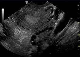
A study led by Durham University’s Fetal and Neonatal Research Lab, UK, took 4D ultrasound scans of 100 pregnant women to see how their unborn babies responded after being exposed to flavours from foods eaten by their mothers.
Researchers looked at how the fetuses reacted to either carrot or kale flavours just a short time after the flavours had been ingested by the mothers.
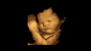
Fetuses exposed to carrot showed more “laughter-face” responses while those exposed to kale showed more “cry-face” responses.
Their findings could further our understanding of the development of human taste and smell receptors.
The researchers also believe that what pregnant women eat might influence babies’ taste preferences after birth and potentially have implications for establishing healthy eating habits.
The study is published in the journal Psychological Science.
pregnant lady/Commons.wikimedia.org
Humans experience flavour through a combination of taste and smell. In fetuses it is thought that this might happen through inhaling and swallowing the amniotic fluid in the womb.
Lead researcher Beyza Ustun, a postgraduate researcher in the Fetal and Neonatal Research Lab, Department of Psychology, Durham University, said: “A number of studies have suggested that babies can taste and smell in the womb, but they are based on post-birth outcomes while our study is the first to see these reactions prior to birth.
“As a result, we think that this repeated exposure to flavours before birth could help to establish food preferences post-birth, which could be important when thinking about messaging around healthy eating and the potential for avoiding ‘food-fussiness’ when weaning.
“It was really amazing to see unborn babies’ reaction to kale or carrot flavours during the scans and share those moments with their parents.”
The research team, which also included scientists from Aston University, Birmingham, UK, and the National Centre for Scientific Research-University of Burgundy, France, scanned the mothers, aged 18 to 40, at both 32 weeks and 36 weeks of pregnancy to see fetal facial reactions to the kale and carrot flavours.
Mothers were given a single capsule containing approximately 400mg of carrot or 400mg kale powder around 20 minutes before each scan. They were asked not to consume any food or flavoured drinks one hour before their scans.
A 4D scan image of a fetus showing a neutral face/CREDIT: FETAP (Fetal Taste Preferences) Study, Fetal and Neonatal Research Lab, Durham University.
The mothers also did not eat or drink anything containing carrot or kale on the day of their scans to control for factors that could affect fetal reactions.
Facial reactions seen in both flavour groups, compared with fetuses in a control group who were not exposed to either flavour, showed that exposure to just a small amount of carrot or kale flavour was enough to stimulate a reaction.
Co-author Professor Nadja Reissland, head of the Fetal and Neonatal Research Lab, Department of Psychology, Durham University, supervised Beyza Ustun’s research. She said: “Previous research conducted in my lab has suggested that 4D ultrasound scans are a way of monitoring fetal reactions to understand how they respond to maternal health behaviours such as smoking, and their mental health including stress, depression, and anxiety.
“This latest study could have important implications for understanding the earliest evidence for fetal abilities to sense and discriminate different flavours and smells from the foods ingested by their mothers.”
Co-author Professor Benoist Schaal, of the National Centre for Scientific Research-University of Burgundy, France, said: “Looking at fetuses’ facial reactions we can assume that a range of chemical stimuli pass through maternal diet into the fetal environment.
“This could have important implications for our understanding of the development of our taste and smell receptors, and related perception and memory.”
The researchers say their findings might also help with information given to mothers about the importance of taste and healthy diets during pregnancy.
They have now begun a follow-up study with the same babies post-birth to see if the influence of flavours they experienced in the womb affects their acceptance of different foods.
Research co-author Professor Jackie Blissett, of Aston University, said: “It could be argued that repeated prenatal flavour exposures may lead to preferences for those flavours experienced postnatally. In other words, exposing the fetus to less ‘liked’ flavours, such as kale, might mean they get used to those flavours in utero.
“The next step is to examine whether fetuses show less ‘negative’ responses to these flavours over time, resulting in greater acceptance of those flavours when babies first taste them outside of the womb.”
Researchers have discovered that some immunotherapy treatments used to treat cancer can cause fertility damage.
It means these treatments could affect the future fertility and hormonal health of female cancer survivors, prompting experts to call for more research and preventative measures, such as freezing eggs.
Led by the Biomedicine Discovery Institute at Monash University and the Peter MacCallum Cancer Centre, the pre-clinical trial showed that immune checkpoint inhibitors, a common type of immunotherapy drug, resulted in permanent damage to mouse ovaries and the eggs stored inside.
cancer/photo:en.wikipedia.org
Traditional cancer therapies, such as chemotherapy and radiotherapy, are already linked to permanent, negative side effects on the ovaries. This can lead to infertility and premature menopause in young girls and women.
Researchers found that checkpoint inhibitor immunotherapy reduced the number and quality of their eggs, interfered with ovulation, and disrupted the fertility cycle.
Until now the potential fertility side effects of immunotherapy, an emerging and increasingly common cancer treatment that stimulates the immune system, have been unknown.
The study found that a type of immunotherapy called immune checkpoint inhibitors, which ‘release the brakes’ on the immune system to enhance a patient’s ability to fight cancer, could impair immediate and future fertility.
Its authors said studies in female patients were now needed to investigate the findings. In the meantime, fertility preservation through egg or embryo freezing should be considered for women using these immunotherapies.
“Initially these treatments were thought to be less damaging (than chemo and radiotherapy) in the context of off-target effects to the body in general,” Ms Alesi said. “However, it is now clear that inflammatory side effects in other organ systems are quite common with these drugs.
“Our study highlights that caution should be exercised by clinicians and their patients, for whom fertility may be a concern. Studies in women receiving these drugs must now be prioritised.”
Peter MacCallum Cancer Centre Specialist Medical Oncologist Professor in breast cancer and a senior author on the study Sherene Loi said further research into how these drugs impact the ovarian function and fertility of women receiving these drugs must be prioritised and should be included in future clinical trials involving women of reproductive age.
“Our study further highlights that fertility discussions are critical for all age appropriate women who are recommended to receive chemotherapy as well as immunotherapy,” Professor Loi said.
“Appropriate interventions that can preserve fertility and ovarian function can be implemented to facilitate pregnancies in the future, post completion of treatment. These interventions need to be implemented in a timely manner, so as not to delay anti-cancer treatment.
“Immunotherapy is now becoming a standard of care for many women with curable early stage breast cancer, due to impressive results in reducing breast cancer recurrences, but further research into the long-term effects of immunotherapy is needed.”
Apart from drugs that block ovaries from producing hormones during chemotherapy, and strategies to prevent premature menopause in younger women, Ms Alesi said egg and embryo freezing was the only fertility preservation measure available.
She said it was important to remember that embryo freezing was expensive, invasive and did not prevent ovarian damage. This meant that premature menopause could still be a risk for these women.
“Therefore, we are now prioritising investigation of targeted ovarian preservation strategies that aim to prevent the damage to the ovary from occurring in the first place, without interfering with the drugs’ ability to fight the cancer” she said.
Babies and children under three years old are less likely to develop croup if their mothers took fish oil and vitamin D supplements during pregnancy, according to new results from a clinical trial.
Croup is a viral chest infection that affects young children. It causes a characteristic ‘barking’ cough, a hoarse voice and difficulty breathing. Croup is common and usually mild, but some children will need hospital treatment and breathing support.
The study was presented by Dr Nicklas Brustad, a clinician and postdoctoral researcher working on the Copenhagen Prospective Studies on Asthma in Childhood (COPSAC) at Copenhagen University Hospital, Denmark.
He told the Congress: “There is currently no vaccine against the pathogen that causes this disease. Therefore, other preventive strategies are needed, and measures initiated during pregnancy might be important since croup occurs in babies and young children. For such purpose, there is evidence that both vitamin D and fish oil could have an influence on the immune system.”
croup-chest infection/photo:en.wikipedia.org
The study included 736 pregnant women being cared for by COPSAC from 2010. The women were divided up into four groups. One group were given a high-dose vitamin D supplement (2800 international units per day) and fish oil containing long-chain n-3-polyunsaturated fatty acids (2.4 grams), the second group were given high-dose vitamin D and olive oil, the third group were given standard-dose vitamin D (400 international units per day) and fish oil, and the final group were given standard-dose vitamin D and olive oil. All the women took the supplements daily from their 24th week of pregnancy until one week after their babies were born. Neither the women nor the researchers knew which supplements they were taking until the end of the study.
Researchers monitored the children until they were three years old and any who were suspected of suffering from croup were diagnosed by a doctor or via their medical records. There was a total of 97 cases of croup amongst the children.
Overall, children whose mothers took the fish oil had an 11% risk of croup, compared to 17% in the children whose mothers took olive oil (a 38% decrease). Children whose mothers took high-dose vitamin D had an 11% risk of croup, compared to an 18% risk in those whose mothers took the standard-dose vitamin D (a 40% decrease).
Dr Brustad said: “Our findings suggest that vitamin D and fish oil could be beneficial against childhood croup in sufficiently high doses. These are relatively cheap supplements meaning that this could be a very cost-effective approach to improving young children’s health.
Vitamin D and fish oil can stimulate the immune system to help babies and young children clear infections more effectively.”
The research team working at COPSAC have already investigated other potential benefits of vitamin D and fish oil during pregnancy, including its effects on bone development, the central nervous system, body composition and asthma. They will continue to follow the children in the study and plan to investigate why some children are more prone to infections in childhood than others.
Professor Rory Morty from the University of Heidelberg is chair of European Respiratory Society’s lung and airway developmental biology group and was not involved in the research. He said: “We know that lung health in babies and young children can be influenced during pregnancy. For example, babies whose mothers smoke tend to have worse lung health. We are increasingly seeing that elements of a mother’s diet can also help or hinder a baby’s lung development.
“This research suggests that taking vitamin D and fish oil supplements during pregnancy could have benefits for babies and young children. We would like to see further research in this area to support these findings as this could lead to new recommendations for supplementation during pregnancy. Pregnant women should always speak to their doctor before taking supplements.”
The language used by doctors when diagnosing female patients with polycystic ovary syndrome (PCOS) can negatively impact their wellbeing and how they view their condition later on in life, new research finds.
PCOS is a condition that affects the working of ovaries and can result in a range of physical symptoms (irregular periods or none at all) and metabolic issues (weight gain). Researchers from the University of Surrey found that the use of the word ‘raised’ by practitioners when discussing test results can lead to higher levels of body dissatisfaction and dieting behaviour amongst women, whilst the use of the word ‘irregular’ can result in concerns about fertility.
Jane Ogden, Professor of Health Psychology at the University of Surrey, said: “Diagnostic consultations may take a few minutes, yet how these minutes are managed, what words are used and how this makes a patient feel may change how they make sense of their condition and influence their wellbeing in the longer term. It is important that doctors have an awareness of the words they use and think about how they could be perceived by patients.”
pregnant lady/Commons.wikimedia.org
In one of the first studies of its kind, researchers from Surrey investigated the impact of PCOS diagnostic consultations and if the language used affected the subsequent wellbeing of patients.
To assess the impact, researchers surveyed 147 females with PCOS and asked about their satisfaction with their consultation, the language used during it and their overall wellbeing.
Researchers found that those who had felt uncomfortable with the consultation process were more likely to report poorer body esteem, reduced quality of life and greater concerns about health in later life. Over a quarter of those surveyed were dissatisfied with how doctors managed their distress and were unhappy with the lack of rapport they had with their practitioners.
“Words matter, as patients often replay conversations that they have had with doctors in a bid to make sense of situations. Although words such as ‘raised’ and ‘irregular’ are simple words they are vague which can cause women to worry, as they automatically think the worst, as they have not been provided with all the facts. Such anxiety at the time of diagnosis, can negatively impact how they feel about themselves as their life progresses,” Professor Ogden added.
Men, unlike women, do not have a menopause to say they cease to help in fertility but a recent study has found that the chance of natural conception can be affected by the age of the male partner, particularly in the genetic health of sperm cells.
Despite a wide belief and celebrity examples of Charlie Chaplin or Luciano Pavarotti, which have kept alive the notion that male fertility goes on forever, the new study in IVF couples shows quite clearly that live birth outcome is clearly affected by the age of the male partner.
“Our study found an independent effect of male age on the cumulative incidence of live birth,” said investigator Dr Laura Dodge from Harvard Medical School, Boston, USA. He will present the study’s results on Tuesday at the 33rd Annual Meeting of ESHRE in Geneva.
The study was an analysis of IVF cycles performed at an IVF centre in Boston between 2000 and 2014. About 19,000 cycles performed in 7,753 couples were analyzed. The female partners were stratified in four age bands: under 30, 30-35 years, 35-40 years, and 40-42. Men were stratified into these same four age bands, with an additional band of 42 and over.
As expected, the cumulative live birth rate was lowest in those couples where the female partner was in the 40-42 age band, and in this group the age of the male partner had no impact. In other bands of female age, the cumulative incidence of live birth was significantly affected by male partner age and was found to decline as the man grew older.
For example, in couples with a female partner aged under 30, a male partner aged 40-42 was associated with a significantly lower cumulative birth rate (46%) than a male partner aged 30-35 (73%). Similarly, in couples with a female partner aged 35-40 years live birth rates were higher with a younger than with an older male partner.
“Generally,” explained Dr Dodge, “we saw no significant decline in cumulative live birth when women had a male partner the same age or younger. Where we see significant decreases in the cumulative incidence of live birth is among women with male partners in the older age bands.”
Dr Dodge noted that in natural conceptions increasing male age is associated with a decreased incidence of pregnancy, increased time to pregnancy, and increased risk of miscarriage. The mechanisms are unclear but may include impaired semen parameters, increased DNA damage in sperm, and epigenetic alterations in sperm that affect fertilisation, implantation, or embryo development, she said.
This is the first study to calculate the cumulative incidence of live birth while jointly stratifying on multiple bands of both male and female age.