How being squeezed contributes to risk of breast cancer cells

A recent study conducted by scientists working in Adelaide University and published in the journal Science Advances has shown the reason as to why certain cancers may grow and survive the body, whereas others do not. It happens that the hard mechanical stress to which the early cancer cells undergo as they are squeezed into a narrow area, causes some of the cancer cells to grow quicker, not to grow, as would otherwise be supposed.

This squeeze worked to the favor of the early breast cancer cells as scientists discovered.

The key point that was explained by the lead researcher, Professor Michael Samuel, of the Centre of Cancer Biology at Adelaide University and the Basil Hetzel Institute is that these breast cancer cells steal a particular sensor – one that our bodies rely on to sense touch – and use it to divide quickly and aid them in making their escape off the major tumour.

The process creates an indefinitely lasting mechanical memory in the breast cancer cells and it still contributes towards aggressive behaviour even after the pressure itself has been removed, Professor Samuel said.

The tumours which are solid are exposed to a lot of physical pressure when the disease is at its early stage of development, as the cancer cells grow in tissues that are limited in space, e.g. the milk ducts of the breast. Up to this day, the mechanism by which these cancer cells detect this pressure and whether or not it impacts the progression of the disease is unknown.

We have a tendency to believe that cancer is a genetic disease, but through this work we know that there is the same importance of physical forces within the tumours as the cause of cancer as there are genetic changes that cause cancer.

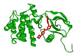
The researchers discovered that cancer cells respond to pressure via a molecule named PIEZO1, which is a hole in the cell that relates the interior of a cell to the exterior environment. Upon pressure stimulation, PIEZO1 enables the movement of calcium ions into the cell and subsequent signal transduction containing the Rho-ROCK pathway – a central regulator of cell movement, shape and growth.

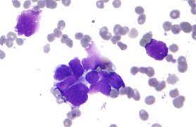
The team demonstrated that mechanical pressure of a short duration that is obtained through compressing cancer tissue was sufficient to cause tumour growth to increase significantly. Mechanically compressed tumours in laboratory models of breast cancer became larger and the cancer cells in them fragmented faster than control groups.

In addition to promoting growth, compression was also identified to drive cancer cells into a more aggressive, invasive, state in a process known as epithelial-mesenchymal transition. When either of the PIEZO1 or the Rho-ROCK pathway had, however, been inhibited with the help of suitable drugs, compression did not propel cancer aggressiveness, making their role in this process definite.

Co-lead author Dr Sarah Boyle mentioned that one of the most significant findings was that the cancer aggressiveness effects of compression remained even after removal of the force itself.

According to Dr Boyle, even relatively short durations of pressure can lead to a mechanical memory by altering the way the DNA is packed into the cell, by chemically modifying the histone proteins.

These changes, which are called epigenetic changes, are modifications of the interpretation of the DNA code by the cell, which enables the process of switching on some genes that promote tumour growth and aggressiveness.

This type of epigenetic mechanical memory offers a molecular basis to the long term effects of short term mechanical forces on the cell level of the behaviour of tumours.

Notably, the research established that PIEZO1 is over-expressed in human breast cancers compared to normal breast tissue, and that the level of PIEZO1 differs among the patients. The high PIEZO1 levels have been linked to low patient survival implying that the identical pressure-detecting system found in test animals would probably be applicable in human cancer.

The results indicate a little-known role of mechanical pressure in the development of cancer aggressiveness and represent the PIEZO1 -Rho-ROCK pathway as a possible new therapeutic objective that can be used as an early intervention.

According to the researchers, future therapies can restrict tumour growth and invasiveness by interfering with the sensory and response of cancer cells to mechanical pressure. The results can also be applied in diagnosing the patients who are susceptible to aggressive breast cancers due to excessively high concentrations of PIEZO1.

That work has opened up a whole new field of so-called mechanotherapy – the use of treatments that disrupt the mechanical signals that tumours are dependent on to develop and spread out, as cancers grow to be mechanically responsive diseases, said Professor Samuel.

Immune targets for chemotherapy-resistant breast cancers identified

Scientists have identified immune cell types that could be targeted to develop specific immunotherapies in chemotherapy-resistant breast cancers.

Researchers from King’s College London and The Institute of Cancer Research, London, with support from Breast Cancer Now, have performed a deep dive into the different immune markers within tumour tissues and blood samples of early breast cancer patients whose cancer failed to respond to chemotherapy given to them prior to surgery.

The research, published today in Clinical Cancer Research, a journal of the American Association for Cancer Research, gives insight into the function of immune cells in patients with chemotherapy-resistant breast cancers. While chemotherapy may not kill cancer cells in these high-risk patients, immunotherapy, a type of treatment that helps the immune system to attack cancer cells, may provide a benefit.

To investigate the immune environment that surrounds these chemotherapy resistant tumours, researchers employed multiple and novel complementary technologies looking at proteins and genes on both pre-treatment and post-treatment breast cancer tissue. They also measured how 1,330 cancer and immune-related genes within cancer tissues were affected by chemotherapy.

Flax seeds help women combat menstrual complications and fight post-menopausal risk of breast cancer, say studies

They found that chemotherapy resistant cancer cells had very few immune cells around them, but chemotherapy did induce changes in several immune cell types. Specifically, they found increases in the number of “innate” (first responder) cells such as neutrophils and natural killer (NK) cells. NK cells help the body to fight infection and cancer. But analysis found the increased NK cells in patients with chemotherapy resistant disease lacked cytotoxic activity – the ‘killing instinct’.

Researchers also found immune-related genes associated with NK cells were those associated with cell inhibition or exhaustion, which meant NK cells were unable to fight cancer cells. This new insight into the behaviour of NK cells could be used to develop specific immunotherapies for these high-risk patients. This would need to be investigated in future clinical trials.

These findings also show that blood monitoring during chemotherapy may help predict chemotherapy response early, potentially allow for tailoring of treatment prior to surgery.

Lead author Dr Sheeba Irshad, Cancer Research UK Clinician Scientist at King’s College London said: “Chemotherapy resistance in aggressive early breast cancers is a major reason why cancer regrows after treatment, contributing significantly to people not surviving their disease. In order to find the right targets for drug developments, it’s important to have a deep understanding of the complex mechanisms that allow some cancer cells to resist treatment, then hide from our immune system to only re-emerge later when they’re harder to eradicate.

“Our work has identified several cell types that would be worth investigating further to understand how they are interacting with the resistant cancer cell and how we can tweak that for our benefit. I am excited to continue to investigate these findings further.”

chemotherapy

Professor Andrew Tutt, Director of the Breast Cancer Now Toby Robins Research Centre at The Institute of Cancer Research, London, and of the Breast Cancer Now Research Unit at King’s College London, said: “Great strides have been made in harnessing immunotherapies to treat several types of cancer, but we need to do better to realise their potential for patients with breast cancer.

“This exciting work advances our understanding of the interaction between cancer cells and the immune system during treatment, and why existing treatments work well for some patients, but not others. I hope this research will help us to enhance the anti-cancer immune response in breast cancer, particularly for patients whose cancer has not responded well to chemotherapy.”

Dr Kotryna Temcinaite, Senior Research Communications Manager at Breast Cancer Now, said: “With an estimated 35,000 people living with incurable secondary (metastatic) breast cancer in the UK, it’s vital we develop smarter, more effective treatments to ensure fewer people hear the devastating news the disease has returned and spread to other parts of the body. This exciting early-stage research, which has been part-funded by Breast Cancer Now, helps to lay the groundwork for discovering a way to target breast cancer cells that resist chemotherapy treatment. We hope by building on these findings, scientists will ultimately be able to develop immunotherapy treatments that may help more people survive breast cancer.

Non-invasive ‘FAST device’ measures the changing size of tumors below the skin

Engineers at the Georgia Institute of Technology and Stanford University have created a small, autonomous device with a stretchable/flexible sensor that can be adhered to the skin to measure the changing size of tumors below. The non-invasive, battery-operated device is sensitive to one-hundredth of a millimeter (10 micrometers) and can beam results to a smartphone app wirelessly in real-time with the press of a button.

In practical terms, the researchers say, their device—dubbed FAST for “Flexible Autonomous Sensor measuring Tumors”—represents a wholly new, fast, inexpensive, hands-free, and accurate way to test the efficacy of cancer drugs. On a grander scale, it could lead to promising new directions in cancer treatment.

Each year researchers test thousands of potential cancer drugs on mice with subcutaneous tumors. Few make it to human patients, and the process for finding new therapies is slow because technologies for measuring tumor regression from drug treatment take weeks to read out a response. The inherent biological variation of tumors, the shortcomings of existing measuring approaches, and the relatively small sample sizes make drug screenings difficult and labor-intensive.

“FAST” sensor/Photo:Stanford University

“In some cases, the tumors under observation must be measured by hand with calipers,” says Alex Abramson, first author of the study and a recent post-doc in the lab of Zhenan Bao at the Stanford School of Engineering and now an assistant professor at Georgia Tech. The use of metal pincer-like calipers to measure soft tissues is not ideal, and radiological approaches cannot deliver the sort of continuous data needed for real-time assessment. FAST can detect changes in tumor volume on the minute-timescale, while caliper and bioluminescence measurements often require weeks-long observation periods to read out changes in tumor size.

FAST’s sensor is composed of a flexible and stretchable skin-like polymer that includes an embedded layer of gold circuitry. This sensor is connected to a small electronic backpack designed by former post-docs and co-authors Yasser Khan and Naoji Matsuhisa. The device measures the strain on the membrane—how much it stretches or shrinks—and transmits that data to a smartphone. Using the FAST backpack, potential therapies that are linked to tumor size regression can quickly and confidently be excluded as ineffective or fast-tracked for further study.

The researchers say that the new device offers few significant advances.

  1. It provides continuous monitoring, as the sensor is physically connected to the mouse/human patients and remains in place over the entire experimental period.
  2. FAST can detect changes in tumor volume on the minute-timescale, while caliper and bioluminescence measurements often require weeks-long observation periods to read out changes in tumor size.
  3. FAST is both autonomous and non-invasive. It is connected to the skin, not unlike a band-aid, battery operated and connected wirelessly. The mouse/human patients are free to move unencumbered by the device or wires, and scientists do not need to actively handle the mice following sensor placement.
  4. FAST packs are also reusable, cost just $60 or so to assemble and can be attached to the mouse/human patients in minutes.
  5. FAST could significantly expedite, automate and lower the cost of the process of screening cancer therapies.

FAST’s sensor is composed of a flexible and stretchable skin-like polymer that includes an embedded layer of gold circuitry.\/Photo:Alex Abramson, Bao Group, Stanford University

The breakthrough is in FAST’s flexible electronic material. Coated on top of the skin-like polymer is a layer of gold, which, when stretched, develops small cracks that change the electrical conductivity of the material. Stretch the material and number of cracks increases, causing the electronic resistance in the sensor to increase as well. When the material contracts, the cracks come back into contact and conductivity improves.

Both Abramson and co-author Naoji Matsuhisa, an associate professor at the University of Tokyo, characterized how these crack propagation and exponential changes in conductivity can be mathematically equated with changes in dimension and volume.

One hurdle the researchers had to overcome was the concern that the sensor itself might compromise measurements by applying undue pressure to the tumor, effectively squeezing it. To circumvent that risk, they carefully matched the mechanical properties of the flexible material to skin itself to make the sensor as pliant and as supple as real skin.

“It is a deceptively simple design,” Abramson says, “But these inherent advantages should be very interesting to the pharmaceutical and oncological communities. FAST could significantly expedite, automate and lower the cost of the process of screening cancer therapies.”

Indian, US, Spain surgeons win global robotic surgery innovation awards

Robotic surgeons from the US, India and Spain were named the top three winners in the KS International Robotic Surgery Innovation competition, as robotic surgery slowly becomes mainstream.

The winners were selected by an international jury form Oxford and Stanford Universities, and New-Delhi based AIIMS, from the fields of urology, gynaecology, general surgery, hepato-biliary-pancreatic surgery, colorectal, head and neck, paediatric and joint replacement surgeries.

The winning entry of Dr Jihad Kaouk, department of urology, Cleveland Clinic in Ohio was titled Single Port Robot-Assisted Kidney Transplantation Extraperitoneal Approach’ in the unique competition organised by Michigan-based robotic surgery evangelist Vattikuti Foundation.

Dr Kaouk and his team modified the technique of robotic kidney transplant developed at the Vattikuti Urology Institute and Medanta Medicity.

Kaouk used a da Vinci single port robot for truly minimally invasive surgery.

Indian, US surgeons win global robotic surgery innovation award

“The early results of patients who had undergone robotic kidney transplant through this technique could go home in only 2 days,” the foundation said in a statement.

For Robotic Infraclavicular Approach for Minimally Invasive Neck Dissection,’ the second award went to Dr Sandeep Nayak, Director, Surgical Oncology, Fortis Cancer Institute, Bengaluru.

Dr Nayak innovated a robotic technique to perform very major cancer surgery of the head neck to clear the lymph nodes in the neck with quick patient recovery and minimal discomfort.

cancer cells/photo:en.wikipedia.org

The third award went to a team of Dr Alberto Piana, Dr Paolo Verri, and Dr Alberto Breda of Oncology Urology and Kidney Transplant Surgery, Fundacio Puigvert, Barcelona, Spain for their entry of 3D Augmented Reality Guided Robotic Assisted Kidney Transplantation’.

The KS National Robotic Surgery Video’ competition is being organised in India since 2015 by Vattikuti Foundation. This year, it went international for the first time.

“As surgeons continue to innovate newer procedures in robotic surgery, the Vattikuti Foundation will continue to invest and make it accessible to other surgeons,” said Raj Vattikuti, president of Vattikuti Foundation.