Understanding which cells within a tumor will go on to form metastases remains one of the major challenges in cancer research. A study led by the Cell Plasticity in Development and Disease laboratory, headed by Ángela Nieto at the Institute for Neurosciences (IN), a joint center of the Spanish National Research Council (CSIC) and Miguel Hernández University (UMH) of Elche, offers an unexpected answer: the cells that will give rise to metastases can already be identified within the primary tumor.
The study, published in Nature Communications, combines the analysis of a mouse model of breast cancer with patient data. The results show that, at the invasive front of the tumor, there is a specific population of cells capable of both invading and either proliferating or entering a dormant state. This balance determines whether cells that escape the tumor can initiate new tumor growths in distant organs, the feared metastases.
Nieto’s team has been studying the epithelial-to-mesenchymal transition (EMT) for decades, a program that controls cell migration during embryonic development and is reactivated in tumors to enable cancer cells to spread and form metastases. In this new study, the researchers go a step further by showing that metastatic ability does not arise randomly and is not exclusively driven by the microenvironment of the target organ. Instead, metastatic potential is already determined within a subset of cells present in the primary tumor, which adopt a highly metastatic state orchestrated by a key factor: the Prrx1 gene.

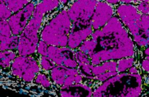
Image of a mouse breast tumor showing distinct cellular populations (cancer cells in pink). Advanced spatial biology technologies enable the simultaneous identification of all cell types present in the tissue.
Credit: Instituto de Neurociencias UMH CSIC
A key regulator of metastasis
According to Raúl Jiménez Castaño, first author of the study, the Prrx1 gene acts as a true master regulator of tumor behavior. “We recently discovered that Prrx1 is crucial for cancer dissemination, and thanks to this work, we now know that it not only activates the programs that allow cells to detach from the primary tumor, but also controls their ability to proliferate and form metastases or enter a dormant state that can last for years”, he explains.
“What is most striking is that the levels of Prrx1 determine its effect on cancer cells”, adds Nieto. This finding helps explain a paradox observed in many tumors: highly invasive cells do not always give rise to metastases, and highly proliferative cells also tend not to spread.
“Without Prrx1, cells do not disseminate; at very high levels, they spread massively but lose the ability to seed and grow in other organs. Only at intermediate levels do they achieve an optimal balance between mobility and growth”, explains the researcher. In these conditions, cells combine invasiveness and proliferation, making them the most dangerous from a clinical perspective.
To reach these conclusions, the team combined mouse genetic models, single-cell analyses, chromatin studies, and spatial transcriptomics techniques that allow researchers to observe the organization and behavior of cells directly within the tumor tissue. The processing and analysis of the large datasets generated from thousands of cells was led by bioinformatics expert from Nieto’s team, researcher Nitin Narwade. In addition, in collaboration with Professor Gema Moreno Bueno from the Universidad Autónoma de Madrid and the MD Anderson Cancer Center Spain Foundation, the researchers analyzed breast cancer patient samples and detected similar patterns of Prrx1 expression, suggesting that the mechanism described could have direct relevance for tumor classification and clinical prognosis.
Taken together, the findings provide new insights into the origin of metastatic potential and open the door to developing strategies to prevent tumor cells from reaching this particularly dangerous state. They also provide a framework for improving patient stratification by identifying markers that predict the risk of metastasis.
Also Read:
Study Links High-Fat Diets To More Aggressive Breast Cancer Behavior
How being squeezed contributes to risk of breast cancer cells