That was the central message of the Manama Declaration, issued at the close of the World Entrepreneurs and Investment Forum (WEIF) held this week in Bahrain.
The declaration places women at the heart of economic transformation, underscoring that empowering them within business and innovation ecosystems is essential to achieving inclusive and sustainable growth.
It also calls for strengthening women’s presence across emerging sectors, including the green, blue and orange economies which respectively promote environmental responsibility, sustainable use of ocean resources and creativity.
Organized by the UN Industrial Development Organization’s (UNIDO) Investment and Technology Promotion Office in Bahrain, the forum convened leaders, investors and entrepreneurs from around the world to shape a more resilient global economy and unlock new opportunities for women-led enterprises.
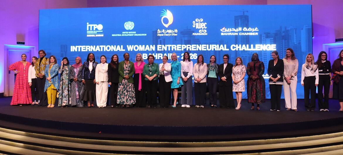
Women entrepreneurs gather on stage at the International Woman Entrepreneurial Challenge 2026 in Manama, Bahrain.
“An amazing opportunity”
For many participants, the forum delivered tangible results.
Doris Martin, CEO of DMartin Consultancy in Bahrain, attended in search of meaningful collaboration and found it.
Through business-to-business (B2B) meetings facilitated during the event, she established partnerships with companies in the United Arab Emirates and Morocco.
“This forum has been effective for me,” she told UN News. “I’ve had B2B collaboration with regional companies through Bahrain and through UNIDO.”
Tosin Arwejulo, CEO of Leadership Excel Consultancy and a Nigerian-American entrepreneur based in Bahrain, described the forum as a “powerful networking platform.”
“I’ve had the opportunity to talk to people from literally every continent,” she said. “It has been an amazing opportunity to connect with like-minded leaders.”
From left to right: Tosin Arwejulo, CEO of Leadership Excel Consultancy and a Nigerian-American entrepreneur based in Bahrain, and Doris Martin, CEO of DMartin Consultancy in Bahrain(In the middle).
Shared challenges, shared solutions
The forum’s impact extended beyond the Arab region.
Ayanthi Gurusinghe, President of the Ceylon Chamber of Women Entrepreneurs in Sri Lanka and representative of the South Asian Women Development Forum, attended alongside delegates from India, Nepal, Bangladesh and Pakistan.
She said participants exchanged valuable lessons, particularly around access to finance, a challenge faced by women entrepreneurs across regions.
“It was a good opportunity for networking, to come together, share views, learn and exchange experiences,” she said, expressing appreciation to UNIDO for creating a space where global connections could flourish.
Ayanthi Gurusinghe, President of the Ceylon Chamber of Women Entrepreneurs in Sri Lanka and representative of the South Asian Women Development Forum, at WEIF 2026 in Bahrain.
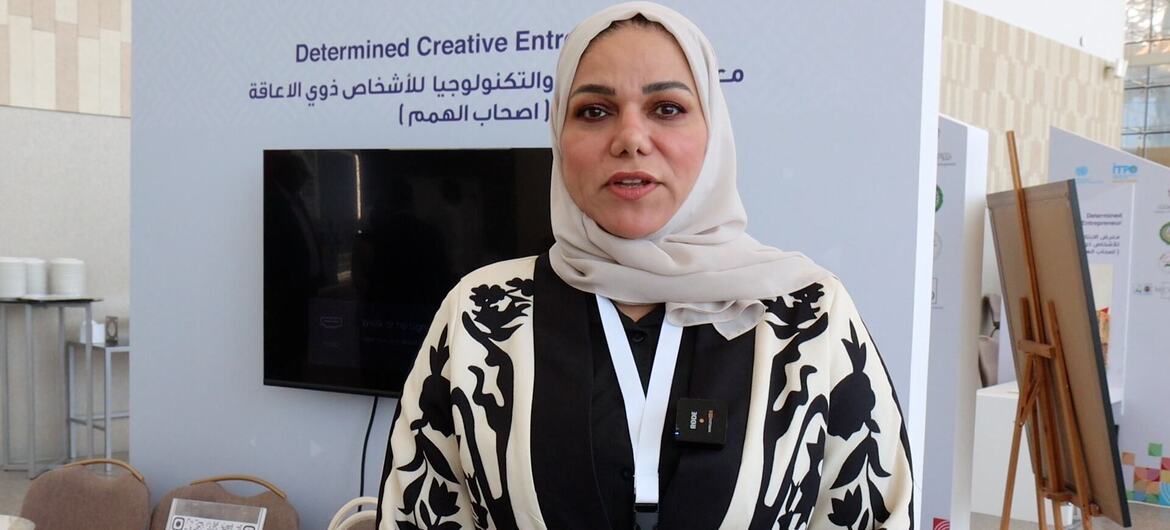
Special focus on artists with disabilities
Beyond panel discussions and networking sessions, the forum also spotlighted inclusive entrepreneurship through three exhibitions including on “Determined Creative Entrepreneur.”
Among the exhibitors was Nisreen Samour of Micro Art Center in Bahrain, which specializes in training and developing artistic talent, with a special focus on artists with disabilities.
“Today, we have about nine students participating, each specializing in an artistic field that aligns with their interests and abilities,” she said.
“I personally train them, and we are currently working on developing their skills and empowering them to produce professional artwork that will help them effectively enter the art market and the job market, thus contributing to increased productivity and the center’s overall performance” she told UN News.
The centre also works with orphans, helping them develop artistic skills and showcase their work publicly — fostering independence and self-reliance through creativity.
Nisreen Samour of Micro Art Center in Bahrain, which specializes in training and developing artistic talent, with a special focus on artists with disabilities.
The role of academia
One recurring theme was the role of higher education in preparing young women to enter the world of entrepreneurship.
In an interview with UN News, Dr. Nihal Al-Najjar, professor at the Royal University for Women in Bahrain, highlighted the need to embed entrepreneurship deeply within academic systems.
“Our academic role is to integrate entrepreneurship not just as a subject, but as a methodology,” she said, explaining that experiential learning and practical application are key. “We encourage students to think, innovate and identify gaps in society, and then work to find solutions.”
She added that universities must go beyond classroom teaching by connecting students to the wider entrepreneurial ecosystem, guiding them towards mentorship, funding opportunities and professional networks when they are ready to launch their own ventures.
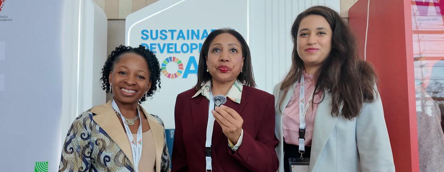
Dina Najar at WEIF2026 in Manama, Bahrain, covering Sustainable Development Goals.
A driving force for sustainable development
The World Entrepreneurs and Investment Forum (WEIF) was preceded by the annual conference of the International Women Entrepreneurial Challenge (IWEC)—a New York-based NGO that helps women-owned businesses to grow.
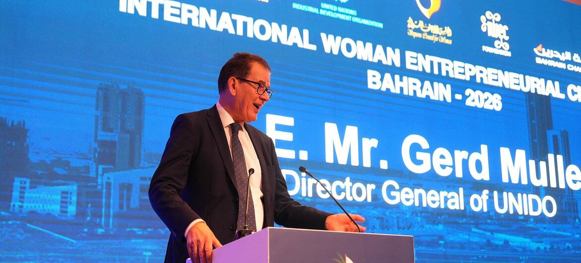
Speaking at the conference, UNIDO Director General Gerd Müller described women’s entrepreneurship as a driving force for sustainable development, global innovation and economic inclusion.
“We must ensure that women everywhere have a level playing field and are given the opportunity to unlock their full potential,” he stated, highlighting persistent barriers to finance, technology, and higher-skilled roles for women globally.
Reaffirming UNIDO’s commitment, he added that “empowering women is essential to achieving the Sustainable Development Goals.” The 17 SDGs, adopted by governments in 2015, provide a blueprint for a more just and equitable future for all people and the planet.
Mr. Müller praised Bahrain’s leadership in supporting entrepreneurship and women’s economic participation and outlined UNIDO’s ongoing work in crisis-affected countries, noting, “UNIDO has just started working to set up recovery programmes in Syria, Sudan, and Palestine… mostly the women there are affected, and they need our support.”
Gerd Müller, Director General of UNIDO, speaking at the International Woman Entrepreneurial Conference in Bahrain 2026.
Women as engines of business
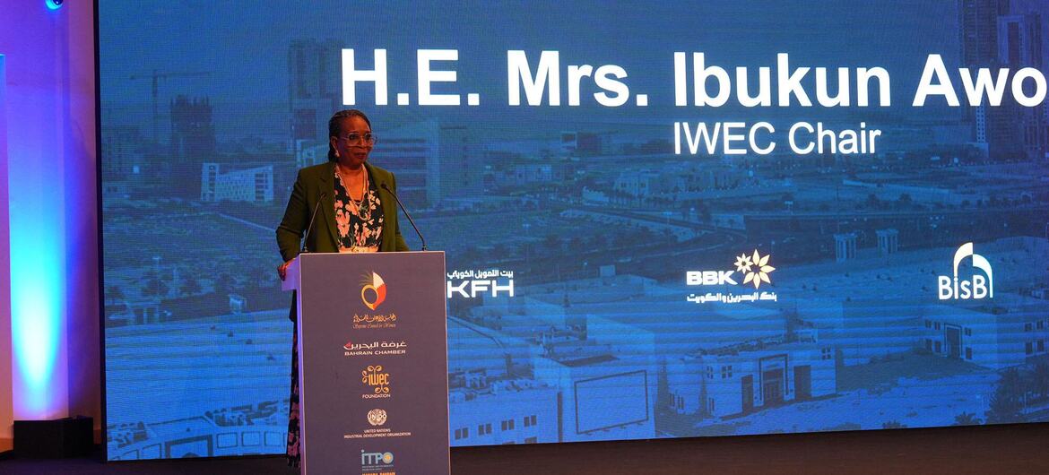
In her opening remarks, IWEC Chair Ibukun Awosika highlighted women’s transformative role as engines of business, leadership, and national progress.
She emphasized that women constitute half of the world’s productive assets and, when empowered, become game changers in corporate and political leadership. Drawing on her own journey, Ms. Awosika recalled building a manufacturing group over 36 years before becoming the first and only woman chair of sub-Saharan Africa’s oldest financial institution, First Bank of Nigeria.
“What women represent to the world is 50 per cent of its assets — productive, constructive assets that are game changers when fully deployed,” she said.
“Every nation that has enough wisdom to invest and to actualize the talent that is in 50 per cent of its population is a country that is set to win.”
H.E. Mrs. Ibukun Awo, IWECC Chair, speaking at WEIF2026 in Bahrain.
Success Stories from Bahrain and Nigeria
According to its website, the International Women Entrepreneurial Challenge (IWEC) has recognized 575 distinguished awardees globally, honoured annually at its conferences and nominated by partner organizations, representing women-owned businesses from across the world.
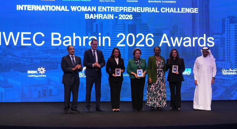
In Bahrain, IWEC recognized a new cohort of awardees from the Middle East and Africa.
Among them was Sonia Mohamed Janahi, founder of Maya La Chocolaterie, who highlighted UNIDO’s role in advancing women’s entrepreneurship.
With UNIDO’s support, Janahi expanded her Bahraini chocolate brand into Africa, establishing a cocoa processing plant in Côte d’Ivoire, creating jobs, promoting ethical supply chains, and ensuring global recognition for African-produced chocolate.
Official event photo from WEIF2026 in Manama, Bahrain.
“UNIDO has played a very vital role in my progression. They have taken my project and opened opportunities for me,” she told UN News, underscoring UNIDO’s commitment to recognizing women entrepreneurs across Bahrain, the Middle East, and Africa.
“This event represents women globally and investments over $6 billion that bring women globally together. It awards women who have not just started a business but have sustained a business and scaled up a business and have had an impact on the economy and on the society.”
Also honoured was Nigerian entrepreneur Oluwakelemi, who discussed her gifting and lifestyle retail business, which employs women and supports household incomes across Nigeria while preparing to expand internationally.
“I am passionate about building scalable African businesses with global reach,” she told UN News. “Not less than 5,000 people have been able to benefit from our business, from our work.”