The battle of the sexes in the egg

The sperm and the egg cell’s nuclei compete for size directly after fertilization and this is necessary for the proper embryonic development. The mouse study with Kobe University participation finally gives meaning to a phenomenon biologists have known for decades.

At the beginning of a new individual’s life, an egg and a sperm fuse to form what biologists call a “zygote,” a fertilized egg. In mammals such as mice and humans, the DNA-carrying nuclei of these cells don’t fuse right away but remain separate as so-called “pronuclei” until just before the first cell division. “This fact has been known for decades, but no one really understood why this separation exists. People just treated it as a descriptive feature rather than something to be tested experimentally,” says Kobe University developmental biologist KYOGOKU Hirohisa. If anything, separate nuclei may increase the risk of errors during the first cell division. In assisted reproduction, embryos in which the maternal and paternal pronuclei are fused early have therefore attracted interest, but these embryos are known to have a lower likelihood of developing to term.

Kyogoku specializes in studying early development directly after fertilization. “The DNA an individual inherits from their mother and father differs not only in its genetic information. It is also known that the maternal genome has a lot of chemical modifications, like little tags, on the molecules around which the DNA is wrapped. The genome from the sperm, on the other hand, has virtually no such modifications. But these proper modifications are essential for embryonic development,” he explains. Thus suspecting that the fusion of pronuclei might have an influence on gene regulation, he teamed up with researchers from RIKEN and, using his special cell manipulation techniques, took a close look at what happened to these modifications when the pronuclei are fused and under other conditions.

In the journal Nature, the team now published that they found that the size of the pronuclei is essential for maintaining the regulatory modifications. Whenever the size of the nucleus was large, either through manipulation or because of the premature fusing of the maternal and paternal pronuclei, the degree of the chemical tagging became low. And when they investigated what keeps the pronuclei’s sizes small in the natural, separated state, they found that the two pronuclei engage in a race to absorb factors that regulate nucleus growth. This then yielded a clear mechanistic explanation for why the developmental potential of fused nuclei is smaller: Because the single nucleus doesn’t have to compete for these factors it ends up much larger and as a result the genome’s regulatory tags get lost. “I find it exciting that a very familiar structure — two separate nuclei in a fertilized egg — turns out to have a clear and functional role, that is, it actively creates a competitive environment inside the cell which helps maintain proper regulation and supports development,” says Kyogoku.

Caption
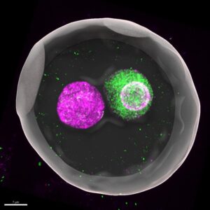
In mammals such as mice and humans, the DNA-carrying nuclei of these cells don’t fuse right away but remain separate as so-called “pronuclei” until just before the first cell division. This 3D-reconstructed image shows a mouse zygote (fertilized egg), where maternal (magenta) and paternal (green) genomes are enclosed in separate pronuclei. “This fact has been known for decades, but no one really understood why this separation exists,” says Kobe University developmental biologist KYOGOKU Hirohisa. Credit:  KYOGOKU Hirohisa

To test their theory, the team temporarily introduced into zygotes with prematurely fused pronuclei an additional pronucleus to re-establish competition. And indeed, this intervention successfully limited nuclear size and partially restored the regulatory marks as well as the developmental potential.

The Kobe University work opens a whole new chapter of research into the very first steps a new life takes. “Even at the beginning of life, spatial organization is not just incidental but fundamentally important,” explains Kobe University biologist Kyogoku. He continues, “This result brings us one step closer to understanding the physical and biological principles that explain why early embryos are both robust and error-prone at the same time.”


Also Read:

Scientists Develop Smart Oxide That Stores Power And Shows Charge Status

Primary Breast Tumours Found To Already Contain Cells With Metastatic Potential

A big step toward producing rhino gametes

To save the northern white rhinoceros from extinction, the BioRescue team is racing to create lab-grown egg and sperm cells of the critically endangered subspecies. The team has now reported a milestone in Science Advances: they have generated primordial germ cells from stem cells – a world’s first.

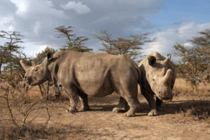
Thirty-three-year-old Najin and her daughter Fatu are the last surviving northern white rhinos on the planet. They live together in a wildlife conservancy in Kenya. With just two females left, this white rhino subspecies is no longer capable of reproduction – at least not on its own. But all hope is not lost: according to a paper published in the journal Science Advances, an international team of researchers has successfully cultivated primordial germ cells (PGCs) – the precursors of rhino eggs and sperm – from embryonic stem cells (ESCs) and induced pluripotent stem cells (iPSCs).


The last two surviving females live in the Ol Pejeta Conservancy in Kenya./CREDIT:Jan Stejskal, Safari Park Dvůr Králové

This represents a major milestone in an ambitious plan. The BioRescue project, which is coordinated by the Leibniz Institute for Zoo and Wildlife Research (Leibniz-IZW) and has been funded by the German Federal Ministry of Education and Research (BMBF) since 2019, wants to save the northern white rhino from extinction. To this end, the scientists are pursuing two strategies – one of them trying to generate viable sperm and eggs from the skin cells of deceased rhinos. The idea is to implant the resulting embryos into closely related southern white rhino females, who will then carry the surrogate offspring to term. And so the northern white rhino subspecies, which humans have already effectively wiped out through poaching, may yet be saved thanks to state-of-the-art stem cell and reproductive technologies.

First success with an endangered species

To get from a piece of skin to a living rhinoceros may be a true feat of cellular engineering, but the process itself is not unprecedented: the study’s co-last author Professor Katsuhiko Hayashi leads research labs at the Japanese universities of Osaka and Kyushu in Fukuoka, where his teams have already accomplished this feat using mice. But for each new species, the individual steps are uncharted territory. In the case of the northern white rhinoceros, Hayashi is working in close cooperation with Dr. Sebastian Diecke’s Pluripotent Stem Cells Technology Platform at the Max Delbrück Center and with reproduction expert Professor Thomas Hildebrandt from Leibniz-IZW. The two Berlin-based scientists are also co-last authors of the current study.

“This is the first time that primordial germ cells of a large, endangered mammalian species have been successfully generated from stem cells,” explains the study’s first author, Masafumi Hayashi of Osaka University. Previously, it has only been achieved in rodents and primates. Unlike in rodents, the researchers have identified the SOX17 gene as a key player in rhinoceros PGC induction. SOX17 also plays an essential role in the development of human germ cells – and thus possibly in those of many mammalian species.

The southern white rhino embryonic stem cells being used in Japan come from the Avantea laboratory in Cremona, Italy, where they were grown by Professor Cesare Galli’s team. The newly derived northern white rhino PGCs, meanwhile, originated from the skin cells of Fatu’s aunt, Nabire, who died in 2015 at Safari Park Dvůr Králové in the Czech Republic. Diecke’s team at the Max Delbrück Center was responsible for converting them into induced pluripotent stem cells.

Next step: cell maturation

Masafumi Hayashi says that they are hoping to use the cutting-edge stem cell technology from Katsuhiko Hayashi’s lab to save other endangered rhino species: “There are five species of rhino, and almost all of them are classified as threatened on the IUCN Red List.” The international team also used stem cells to grow PGCs of the southern white rhino, which has a global population of around 20,000 individuals. In addition, the researchers were able to identify two specific markers, CD9 and ITGA6, that were expressed on the surface of the progenitor cells of both white rhino subspecies. “Going forward, these markers will help us detect and isolate PGCs that have already emerged in a group of pluripotent stem cells,” Hayashi explains.

The BioRescue scientists must now move on to the next difficult task: maturing the PGCs in the laboratory to turn them into functional egg and sperm cells. “The primordial cells are relatively small compared to matured germ cells and, most importantly, still have a double set of chromosomes,” explains Dr. Vera Zywitza from Diecke’s research group, who was also involved in the study. “We therefore have to find suitable conditions under which the cells will grow and divide their chromosome set in half.”

Genetic variation is key for conservation

Leibniz-IZW researcher Hildebrandt is also pursuing a complementary strategy. He wants to obtain egg cells from 22-year-old Fatu and fertilize them in Galli’s lab in Italy using frozen sperm collected from four now deceased northern white rhino bulls. This sperm is thawed and injected into the egg in a process known as intracytoplasmic sperm injection (ICSI). However, Hildebrandt explains that Fatu is not able to bear her own offspring, as she has problems with her Achilles tendons and cannot carry any additional weight. Her mother Najin, meanwhile, is past child-bearing age and also suffers from ovarian tumors. “And in any case, since we only have one donor of natural eggs left, the genetic variation of any resulting offspring would be too small to create a viable population,” he adds.

The team’s top priority, therefore, is turning the PGCs they now have at their disposal into egg cells. “In mice, we found that the presence of ovarian tissue was important in this crucial step,” Zywitza explains. “Since we cannot simply extract this tissue from the two female rhinos, we will probably have to grow this from stem cells as well.” The scientist is hopeful, however, that ovarian tissue from horses could come in useful, as horses are among the rhinos’ closest living relatives from an evolutionary standpoint. If only humans had taken as good care of the wild rhino as they had of the domesticated horse, the immense challenge now facing the BioRescue scientists could perhaps have been avoided altogether.

Also Read:

VMC is biomarker of ageing for nematode; what is its role in Humans?

Clarifying the chaos of narwhals behavior; what are narwhals, how they help [Details]

Sound reveals ‘Ocean giants’ dance with wind to find food

These blood groups linked to your risk of stroke before age 60: Study

Gene variants associated with a person’s blood type may be linked to their risk of early stroke, according to a new meta-analysis published in the latest online issue of Neurology.

Non-O blood types have previously been linked to a risk of early stroke, but the new findings by University of Maryland School of Medicine study showed a stronger link between these blood types with early stroke compared to late stroke, and in linking risk mostly to blood type A.

Study author Braxton D. Mitchell of University of Maryland School of Medicine in Baltimore said, “Specifically, our meta-analysis suggests that gene variants tied to blood types A and O represent nearly all of those genetically linked with early stroke. People with these gene variants may be more likely to develop blood clots, which can lead to stroke.”

The meta-analysis involved a review of 48 studies on genetics and ischemic stroke from North America, Europe and Asia. The studies included 16,927 people with stroke and 576,353 people who did not have a stroke. Of those with stroke, 5,825 people had early onset stroke and 9,269 people had late onset stroke. Early onset stroke was defined as an ischemic stroke occurring before age 60 and late onset stroke was older than 60.

Researchers looked across all the chromosomes to identify genetic variants associated with stroke. They found a link between early stroke and the area of the chromosome that includes the gene that determines A, AB, B or O blood type.

They then divided participants into A, AB, B and O blood types. They compared the prevalence of those blood types in people with early stroke, late stroke and people who did not have a stroke.

Researchers found that people with early stroke were more likely to have blood type A and less likely to have blood type O compared to people with late stroke and people without stroke. Both early and late stroke were also more likely to have blood type B compared to controls.

Blood types/photo: en.wikipedia.org

When looking at people of European ancestry and comparing 5,825 people with early stroke to 29,320 people who did not have a stroke, the meta-analysis found that 48% of people with early stroke had blood type A compared to 45% of people with late stroke and 44% of people without stroke. They also found 35% of people with early stroke had blood type O compared to 39% of those with late stroke and 41% of people without stroke.

After adjusting for sex and other factors, researchers found those who had blood type A had an 16% higher risk of having an early stroke than people with other blood types. Those who had blood type O had a 12% lower risk of having a stroke than people with other blood types.