New ecology tools predict disease transmission among wildlife, humans

The rate that emerging wildlife diseases infect humans has steadily increased over the last three decades. Viruses, such as the global coronavirus pandemic and recent monkeypox outbreak, have heightened the urgent need for disease ecology tools to forecast when and where disease outbreaks are likely.

A University of South Florida assistant professor helped develop a methodology that will do just that – predict disease transmission from wildlife to humans, from one wildlife species to another and determine who is at risk of infection.

The methodology is a machine-learning approach that identifies the influence of variables, such as location and climate, on known pathogens. Using only small amounts of information, the system is able to identify community hot spots at risk of infection on both global and local scales.
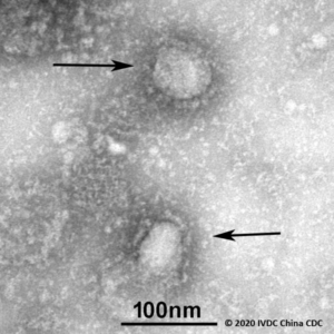
coronavirus

coronavirus

“Our main goal is to develop this tool for preventive measures,” said co-principal investigator Diego Santiago-Alarcon, a USF assistant professor of integrative biology. “It’s difficult to have an all-purpose methodology that can be used to predict infections across all the diverse parasite systems, but with this research, we contribute to achieving that goal.”

With help from researchers at the Universiad Veracruzana and Instituto de Ecologia, located in Mexico, Santiago-Alarcon examined three host-pathogen systems – avian malaria, birds with West Nile virus and bats with coronavirus – to test the reliability and accuracy of the models generated by the methodology.

The team found that for the three systems, the species most frequently infected was not necessarily the most susceptible to the disease. To better pinpoint hosts with higher risk of infection, it was important to identify relevant factors, such as climate and evolutionary relationships.

By integrating geographic, environmental and evolutionary development variables, the researchers identified host species that have previously not been recorded as infected by the parasite under study, providing a way to identify susceptible species and eventually mitigate pathogen risk.

“We feel confident that the methodology is successful, and it can be applied widely to many host-pathogen systems,” Santiago-Alarcon said. “We now enter into a phase of improvement and refinement.”

The results, published in the Proceedings of the National Academy of Sciences, prove the methodology is able to provide reliable global predictions for the studied host–pathogen systems, even when using a small amount of information. This new approach will help direct infectious disease surveillance and field efforts, providing a cost-effective strategy to better determine where to invest limited disease resources.

Bats

Bats/wikipedia

Predicting what kind of pathogen will produce the next medical or veterinary infection is challenging, but necessary. As the rate of human impact on natural environments increases, opportunity for novel diseases will continue to rise.

“Humanity, and indeed biodiversity in general, are experiencing more and more infectious disease challenges as a result of our incursion and destruction of the natural order worldwide through things like deforestation, global trade and climate change,” said Andrés Lira-Noriega, research fellow at the Instituto de Ecologia. “This imposes the need of having tools like the one we are publishing to help us predict where new threats in terms of new pathogens and their reservoirs may occur or arise.”

The team plans to continue their research to further test the methodology on additional host-pathogen systems and extend the study of disease transmission to predict future outbreaks. The goal is to make the tool easily accessible through an app for the scientific community by the end of 2022.

Manuka Honey emerges miracle drug for lung infection if combined with widely used ‘amikacin’

A potential new treatment combining natural manuka honey with a widely used drug has been developed by scientists at Aston University to treat a potentially lethal lung infection and greatly reduce side effects of one of the current drugs used for its treatment.

Manuka honey can also be used to help treat wounds, injuries, improve oral health, soothe a sore throat and treat ulcers. The findings are published in the journal Microbiology.

The scientists in the Mycobacterial Research Group in the College of Health and Life Sciences at Aston University were able to combine manuka honey and the drug amikacin in a lab-based nebulisation formulation to treat the harmful bacterial lung infection Mycobacterium abscessus, said lead author and PhD researcher Victoria Nolan.

Manuka honey is long known to have wide ranging medicinal properties, but more recently has been identified for its broad spectrum antimicrobial activity. Now scientists have found that manuka honey has the potential to kill a number of drug resistant bacterial infections such as Mycobacterium abscessus – which usually affects patients with cystic fibrosis (CF) or bronchiectasis.

Manuka honey could help to clear deadly drug-resistant lung infection – research/Photo:Microbiology Society

According to the Cystic Fibrosis Trust, CF is a genetic condition affecting around 10,800 people – one in every 2,500 babies born in the UK – and there are more than 100,000 people with the condition worldwide. The NHS defines bronchiectasis  as a long-term condition where the airways of the lungs become widened, leading to a build-up of excess mucus that can make the lungs more vulnerable to infection..

In the study, the researchers used samples of the bacteria Mycobacterium abscessus taken from 16 infected CF patients. They then tested the antibiotic amikacin, combined with manuka honey, to discover what dosage was required to kill the bacteria.

Dr Jonathan Cox, senior lecturer in microbiology, Aston University said: “By combining a totally natural ingredient such as manuka honey with amikacin, one of the most important yet toxic drugs used for treating Mycobacterium abscessus, we have found a way to potentially kill off these bacteria with eight times less drug than before.”

As part of the study the team used a lab-based lung model and nebuliser – a device that produces a fine spray of liquid often used for inhaling a medicinal drug. By nebulising manuka honey and amikacin together, it was found they could improve bacterial clearance, even when using lower doses of amikacin, which would result in less life-changing side-effects to the patient.

In the UK, of the 10,800 people living with CF, Mycobacterium abscessus infects 13% of all patients with the condition. This new approach is advantageous not only because it has the potential to kill off a highly drug resistant infection, but because of the reduced side effects, benefitting quality of life and greatly improving survival chances for infected CF patients.

Mycobacterium abscessus is a bacterial pathogen from the same family that causes tuberculosis, but this bug differs by causing serious lung infections in people (particularly children) with pre-existing lung conditions, such as CF and bronchiectasis, as well as causing skin and soft tissue infections. The bacteria is also highly drug resistant.

Currently, patients are given a cocktail of antibiotics, consisting of 12 months or more of antimicrobial chemotherapy and often doesn’t result in a cure. The dosage of amikacin usually used on a patient to kill the infection is 16 micrograms per millilitre. But the researchers found that the new combination using manuka honey, required a dosage of just 2 micrograms per millitre of amikacin – resulting in a one eighth reduction in the dosage of the drug.

Until now Mycobacterium abscessus has been virtually impossible to eradicate in people with cystic fibrosis. It can also be deadly if the patient requires a lung transplant because they are not eligible for surgery if the infection is present.


Medieval friars were ‘riddled with parasites’, new findings reveal

A new analysis of remains from medieval Cambridge shows that local Augustinian friars were almost twice as likely as the city’s general population to be infected by intestinal parasites.

This is despite most Augustinian monasteries of the period having latrine blocks and hand-washing facilities, unlike the houses of ordinary working people.

Researchers from the University of Cambridge’s Department of Archaeology say the difference in parasitic infection may be down to monks manuring crops in friary gardens with their own faeces, or purchasing fertiliser containing human or pig excrement.

The study, published today in the International Journal of Paleopathology, is the first to compare parasite prevalence in people from the same medieval community who were living different lifestyles, and so might have differed in their infection risk.

The population of medieval Cambridge consisted of residents of monasteries, friaries and nunneries of various major Christian orders, along with merchants, traders, craftsmen, labourers, farmers, and staff and students at the early university.

Cambridge archaeologists investigated samples of soil taken from around the pelvises of adult remains from the former cemetery of All Saints by the Castle parish church, as well as from the grounds where the city’s Augustinian Friary once stood.

Most of the parish church burials date from the 12-14th century, and those interred within were primarily of a lower socio-economic status, mainly agricultural workers.

The Augustinian friary in Cambridge was an international study house, known as a studium generale, where clergy from across Britain and Europe would come to read manuscripts. It was founded in the 1280s and lasted until 1538 before suffering the fate of most English monasteries: closed or destroyed as part of Henry VIII’s break with the Roman Church.

The researchers tested 19 monks from the friary grounds and 25 locals from All Saints cemetery, and found that 11 of the friars (58%) were infected by worms, compared with just eight of the general townspeople (32%).

They say these rates are likely the minimum, and that actual numbers of infections would have been higher, but some traces of worm eggs in the pelvic sediment would have been destroyed over time by fungi and insects.

The 32% prevalence of parasites among townspeople is in line with studies of medieval burials in other European countries, suggesting this is not particularly low – but rather the infection rates in the monastery were remarkably high.

“The friars of medieval Cambridge appear to have been riddled with parasites,” said study lead author Dr Piers Mitchell from Cambridge’s Department of Archaeology. “This is the first time anyone has attempted to work out how common parasites were in people following different lifestyles in the same medieval town.”

Cambridge researcher Tianyi Wang, who did the microscopy to spot the parasite eggs, said: “Roundworm was the most common infection, but we found evidence for whipworm infection as well. These are both spread by poor sanitation.”

Standard sanitation in medieval towns relied on the cesspit toilet: holes in the ground used for faeces and household waste. In monasteries, however, running water systems were a common feature – including to rinse out the latrine – although that has yet to be confirmed at the Cambridge site, which is only partly excavated.

Not all people buried in Augustinian friaries were actually clergy, as wealthy people from the town could pay to be interred there. However, the team could tell which graves belonged to friars from the remains of their clothing.

“The friars were buried wearing the belts they wore as standard clothing of the order, and we could see the metal buckles at excavation,” said co-author Craig Cessford of the Cambridge Archaeological Unit.

As roundworm and whipworm are spread by poor sanitation, researchers argue that the difference in infection rates between the friars and the general population must have been due to how each group dealt with their human waste.

“One possibility is that the friars manured their vegetable gardens with human faeces, not unusual in the medieval period, and this may have led to repeated infection with the worms,” said Mitchell.

Medieval records reveal how Cambridge residents may have understood parasites such as roundworm and whipworm. John Stockton, a medical practitioner in Cambridge who died in 1361, left a manuscript to Peterhouse college that included a section on De Lumbricis (‘on worms’).

It notes that intestinal worms are generated by excess of various kinds of phlegm: “Long round worms form from an excess of salt phlegm, short round worms from sour phlegm, while short and broad worms came from natural or sweet phlegm.”

The text prescribes “bitter medicinal plants” such as aloe and wormwood, but recommends they are disguised with “honey or other sweet things” to help the medicine go down.

Another text – Tabula medicine – found favour with leading Cambridge doctors of the 15th century, and suggests remedies as recommended by individual Franciscan monks, such as Symon Welles, who advocated mixing a powder made from moles into a curative drink.

Overall, those buried in medieval England’s monasteries had lived longer than those in parish cemeteries, according to previous research, perhaps due to a more nourishing diet, a luxury of wealth.

‘Increased risk’ donor organs a tough sell to transplant patients

Increasingly, transplant surgeons must initiate a tough conversation: explaining to patients what it means to accept an organ from a person who died from a drug overdose.

Organ donors who recently injected drugs, as well those who were incarcerated or had sex for drugs or money, are among a growing group of people classified as being at “increased risk” of an infectious disease such as hepatitis C or HIV.

But the label may not be doing patients any favors, according to a study about transplant trends and organ discard.

National organ transplant numbers show the United States Public Health Service’s increased risk of infection label (PHS-IR) is associated with hundreds of available organs going unused each year.

Despite the very low risk of disease transmission, patients in need are saying no to these organs, which account for 1 in every 5 deceased donor organs today.

Better guidance is needed for surgeons and patients on how much risk an increased-risk label actually presents to patients, says study author Daniel Kaul, M.D., director of the Transplant Infectious Disease Service at Michigan Medicine.

For a study published in Transplantation, Kaul led an analysis of Organ Procurement and Transplantation Network (OPTN) data showing organs labeled as increased risk are 7 percent less likely to be used than organs without the label.

“Overall utilization was less despite the extraordinarily low risk of disease transmission,” says Kaul, a professor of internal medicine at the University of Michigan. “The organ may have gone to the next person on the transplant waiting list, but it might not have been used at all.”

Also troubling, utilization rates of PHS-IR organs varied dramatically by geography. Depending on the donation service area, transplantation of available adult kidneys from increased-risk donors ranged from 20 to 100 percent, the analysis found.

“What that tells us is there may be a different understanding of true risk associated with this label — from one center to another and even within a center, from one organ specialty to another,” Kaul says.

If the increased-risk label did not exist, an estimated 313 more transplants could be performed in the United States each year, according to the study.

All organs identified as increased risk were less likely to be used with the exception of livers, for which transplant rates were nearly identical.

A possible explanation? Patients with liver failure may be too sick to say no. By comparison, a patient needing a kidney may continue dialysis and wait for another organ offer.

Favorable Odds

For most patients with end-stage organ failure, the immediate risk of dying is greater than the risk of getting an organ with an infection.

After rigorous screening, the risk of HIV or hepatitis C transmission from organ donation is low, at less than 1 percent — much less, for example, than the lifetime chances of dying in a motor vehicle accident.

Co-authored by Michael Volk, M.D., a former U-M physician now practicing in Loma Linda, California, and others, the study was published in a special issue of Transplantation that looked at reducing organ discard while safely maximizing organ availability.

Researchers analyzed criteria of donors from 2010 to 2013. The rate of nonutilization of increased-risk organs, the authors write, is likely now even higher because of a 2015 change to the PHS-IR criteria. Combined with the opioid epidemic, the change resulted in an increase in the proportion of organs with this label from 12 percent to 20 percent.

With little guidance about magnitude of risk, the stigma surrounding drug addiction can lead someone to turn down an organ that could save his or her life.

As of early October, about 116,500 Americans are waiting for an organ transplant.